 Blogs
Blogs
Drawing on the insights of our leadership team, we’ve compiled a global perspective on the state of the bioanalytical industry in 2024. Through thoughtful discussions with our CEO, John Bucksath, and key team members Amy Mize, Mouhssin Oufir, and Brian Wile, KCAS Bio delivers a…
 Blogs
Blogs
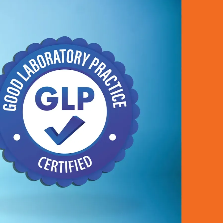
The Importance of GLP Compliance Certificate for Bioanalytical CROs We are proud to celebrate the one-year anniversary of our Good Laboratory Practice (GLP) Test Facility based in Lyon (France), dedicated to…
 Blogs
Blogs
Intracellular cytokine staining (ICS) is a functional immunology assay that uses flow cytometry to assess cytokine production by individual cells. Cytokines play a critical role in developing both physiologically appropriate and pathological immune responses. Interferon-γ (IFN-γ) is an excellent example of this, where it has a key role in…
 Blogs
Blogs
Oligonucleotide therapeutics have recently gained popularity as noted by recent increases in regulatory approvals of oligonucleotides as drugs, novel liquid nanoparticle delivery approaches, and on-target specificity. In the development of bioanalytical assays, there are many challenging aspects to consider when quantitating oligonucleotides, such as non-specific binding,…
 Blogs
Blogs
KCAS Bio has embarked on a global initiative to harmonize its spectral flow cytometry services across four labs in three countries, collaborating with Crux Biolabs and Cytek Biosciences. This effort ensures standardized flow cytometry services across sites in the U.S., Europe, and Australia. As trials increasingly span multiple regions,…
 Blogs
Blogs
We are thrilled to announce a major milestone in the evolution of our global clinical support services. After months of detailed planning, collaboration, and execution, we’ve successfully integrated spectral flow cytometry at our KCAS Bio sites in Philadelphia, PA, USA; Lyon, France; and Crux Biolabs,…
 Blogs
Blogs
Clinical sample kitting plays a vital role in drug development projects. It requires meticulous attention to detail and the experience necessary to ensure that each kit is crafted according to precise specifications. A well-organized clinical kit can be the difference between smooth operations and costly delays. At KCAS…
 Blogs
Blogs
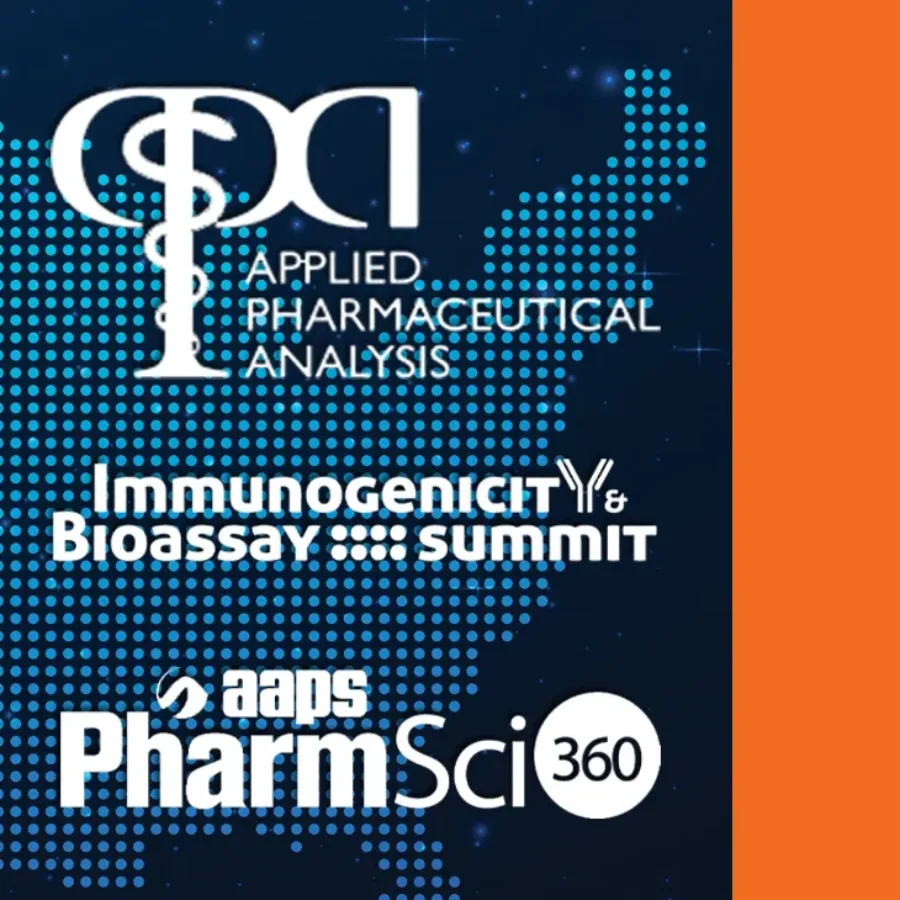
KCAS Bio is excited to announce our participation in several premier conferences throughout the United States this fall. These events bring together experts, innovators, and leaders in pharmaceuticals, biotechnology, and clinical research to exchange insights, explore the latest technologies, and drive the future of scientific discovery. As a trusted…
 Blogs
Blogs
When you examine how to develop and validate/qualify a Biodistribution assay by PCR…
 Blogs
Blogs
KCAS Bio is excited to announce our participation in several key conferences throughout the world. These events present invaluable opportunities for collaboration, innovation, and the sharing of industry insights. As a leading provider of bioanalytical services, KCAS Bio is committed to engaging with thought leaders, scientists, and professionals worldwide…
 Blogs
Blogs
What is the Role of Pharmacokinetics (PK) in Drug Development? Pharmacokinetics (PK) is defined as the study of the fate of a new therapeutic entity (NTE) after it is administered into the body of animals or humans. It involves understanding how the drug is absorbed, distributed, metabolized, and eliminated (ADME).
 Blogs
Blogs
The exploration of Targeted Protein Degraders (TPD) as a therapeutic modality began over twenty years ago. Since then, research and development in this field have steadily advanced. Starting in 2019, rationally designed small molecule TPDs entered clinical trials. This innovative therapeutic modality utilizes the cells’ endogenous protein degradation machinery…