 Blogs
Blogs
From initial receipt to final disposition, sample integrity, traceability, and compliance are the foundation of our bioanalytical operations. We’re taking you behind the scenes to follow the journey of a sample through our facility. Here’s a detailed look at each stage in the lifecycle of a study sample at…
 Podcasts
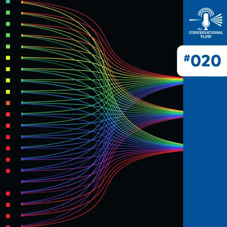
In this milestone 20th episode of “The Conversational Flow” podcast, hosts Adam and Brian dive into impactful developments across oncology, regulatory science, and bioanalytical innovation. The episode begins with a highlight of a recent major Keytruda study showing the first significant improvement in head and neck squamous cell carcinoma…
 Blogs
Blogs
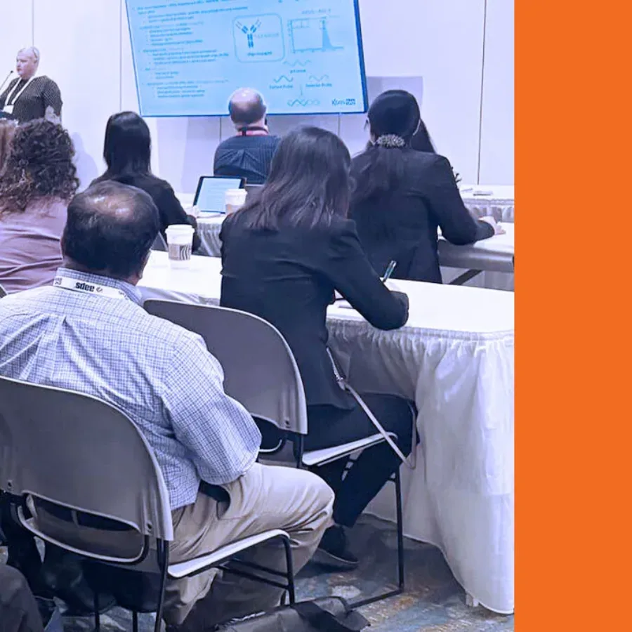
KCAS Bio participated in the AAPS National Biotechnology Conference (NBC), held from May 4 to May 7, 2025, in Boston. It was a productive event that allowed us to connect with sponsors, exchange ideas with peers, and present our latest work in bioanalytical science.
 Posters & Papers
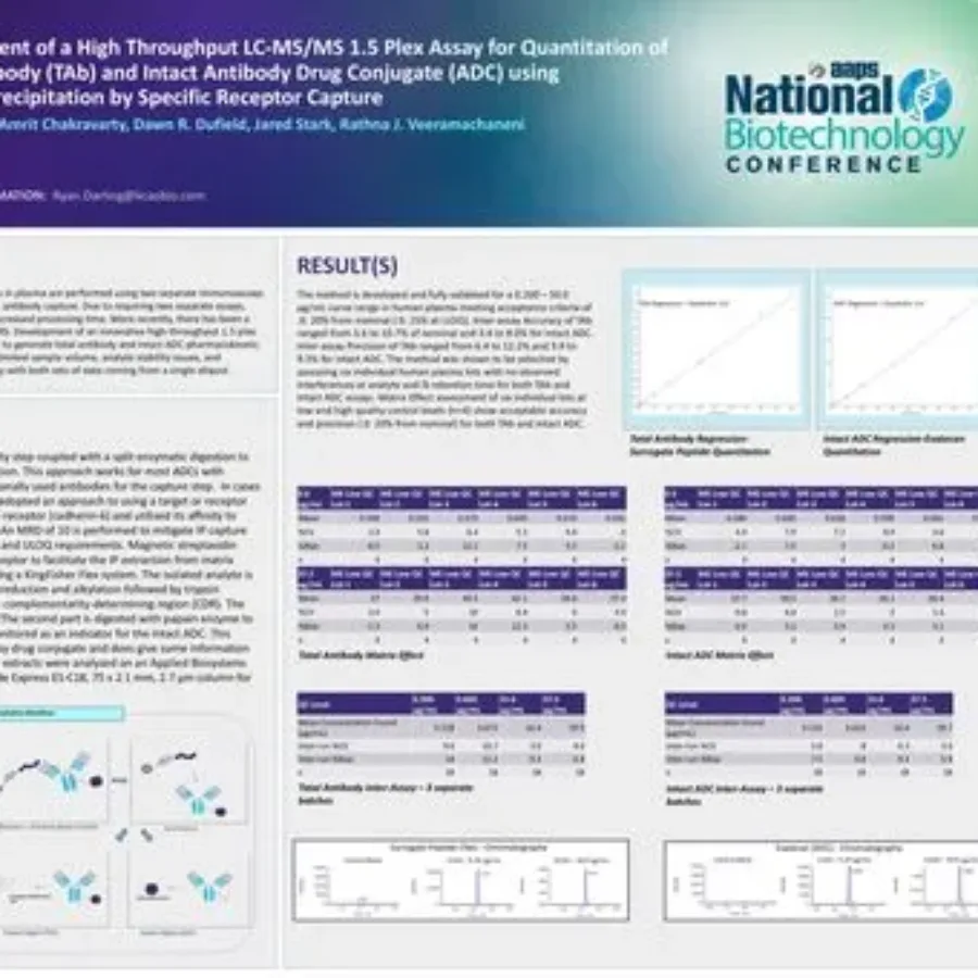
Discover in this poster presented by Ryan Darling at the AAPS National Biotechnology Conference 2025 on the use of a hybrid LC-MS/MS approach for the quantitation of total antibody TAb and intact Antibody Drug Conjugate (ADC). If you have any questions about these services or any…
 Blogs
Blogs
In drug development, the journey from target identification to clinical candidate selection involves a series of critical steps, including compound screening, lead optimization, and pre-clinical testing. Each stage helps narrow the pool of potential therapeutics, with the goal of identifying the most promising candidates for clinical evaluation. Given the…
 Blogs
Blogs
At KCAS Bio, we offer expert flow cytometry services designed to support the evolving bioanalytical needs of immunology and biomarker projects for drug development. Whether you’re working in preclinical or clinical settings, our validated off-the-shelf panels save time, reduce risk, and deliver high-quality, reproducible data you can trust. What…
 Podcasts
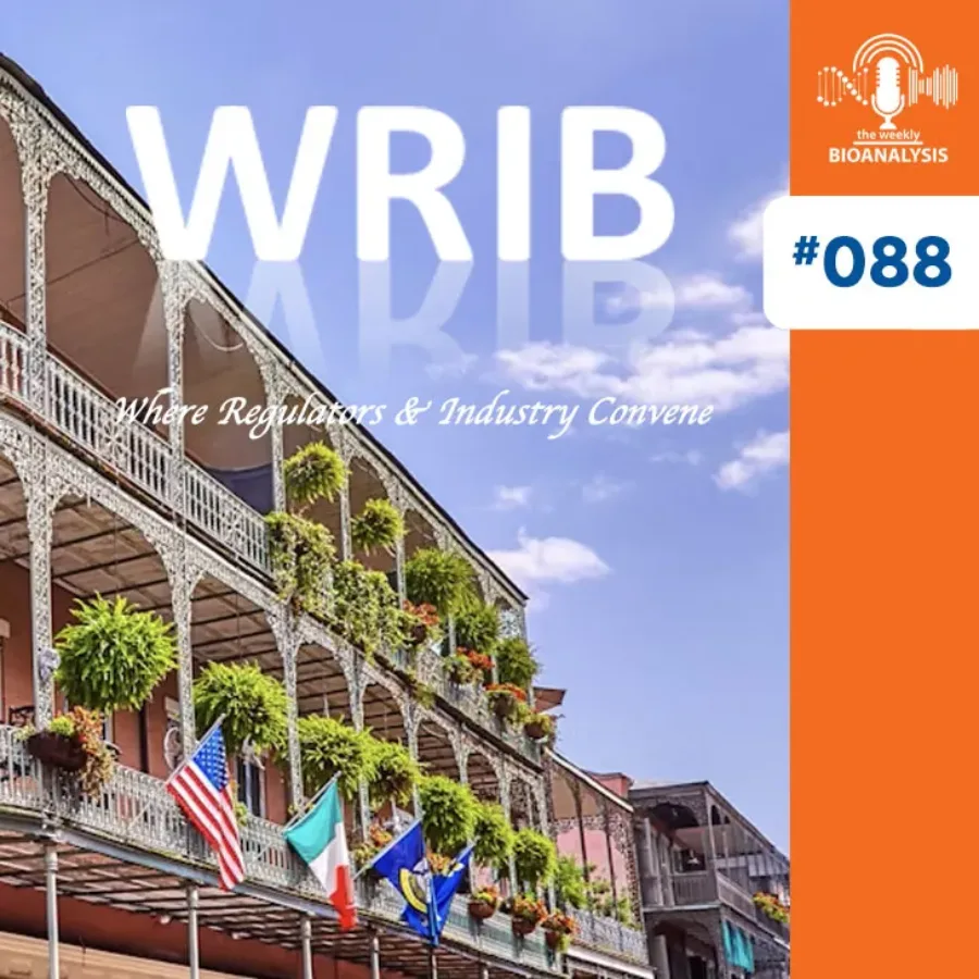
In Episode 88 of The Weekly Bioanalysis, hosts Dominic Warrino, Ph.D. and John Perkins, Ph.D present a special WRIB 2025 recap featuring several KCAS Bio scientists who share firsthand insights from their presentations at the major bioanalytical conference. David Ambrose highlights KCAS Bio’s global harmonization of spectral flow cytometry instruments…
 Blogs
Blogs
KCAS Bio offers a wide range of biomarker services, from cell-based to soluble biomarker analysis, including ligand binding assays (LBA), across a variety of matrix types. Soluble biomarker analysis can be achieved on multiple platforms depending on factors such as sample type, required sensitivity, and whether multiplexing…
 Podcasts
In the 19th episode of “The Conversational Flow”, our hosts, Brian and Adam, dive into the utility and innovation behind backbone panels in spectral flow cytometry, spotlighting a recent paper from BMS that presents a robust, flexible assay designed for immune monitoring using the Aurora platform. They discuss how…
 Posters & Papers
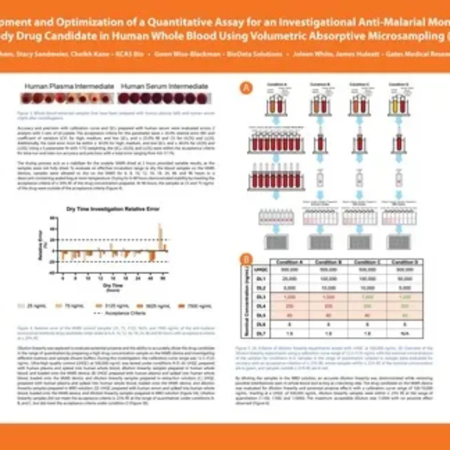
Discover in this poster presented by Jessica Pham at WRIB 2025 on the “Development and Optimization of a Quantitative Assay for an Investigational Anti-Malarial Monoclonal Antibody Drug Candidate in Human Whole Blood Using Volumetric Absorptive Microsampling (VAMS)”. If you have any questions about these services or any others…
 Posters & Papers
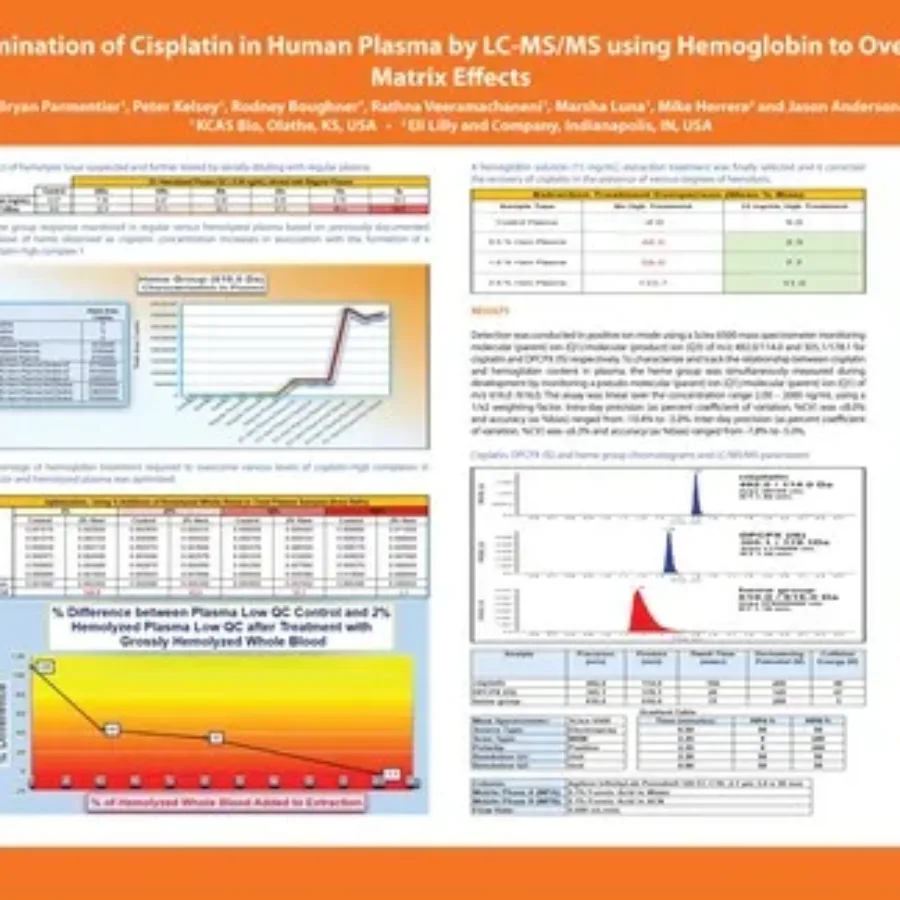
Discover in this poster presented by Bryan Parmentier at WRIB 2025, our work for the sponsor Lilly on the “Determination of Cisplatin in Human Plasma by LC-MS/MS using Hemoglobin to Overcome Matrix Effects“. If you have any questions about these services or any others offered by KCAS Bio,…
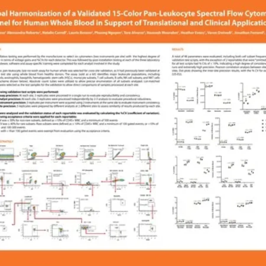 Posters & Papers
Discover in this poster presented by David Ambrose at WRIB 2025, our work on the Global Harmonization of a Validated 15-Color Pan-Leukocyte Spectral Flow Cytometry Panel for Human Whole Blood in Support of Translational and Clinical Applications. If you have any questions about these services or any others…