 Posters & Papers
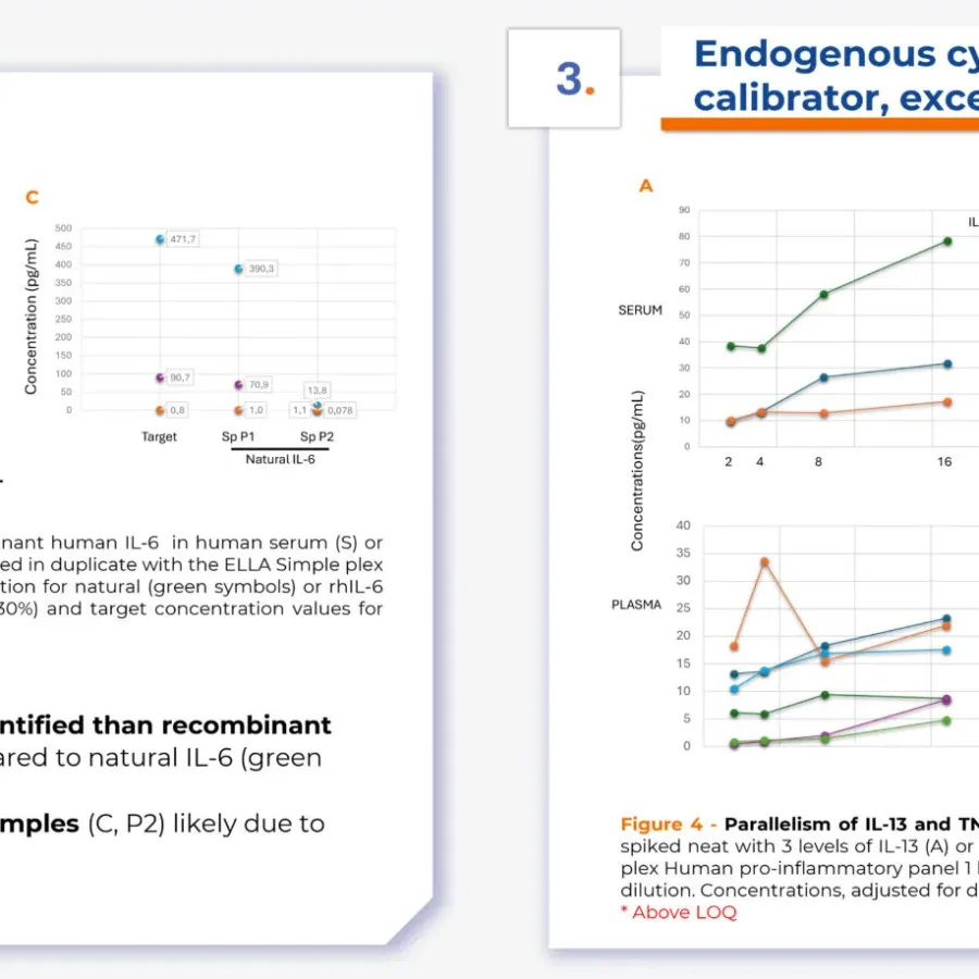
Discover innovative insights into bioanalytical method development with our poster, presented by Christine Bain, Ph.D., at ECCO 2025. This work explores the creation and application of reference samples containing endogenous immune mediators—such as cytokines and chemokines—to effectively evaluate the performance of bioanalytical methods. Download the poster now to learn more…
 Blogs
Blogs
Modern immunology research requires high-complexity panels available wherever the patients are located. Deploying advanced flow cytometry instruments and panels across multiple global sites is therefore a transformative step in harmonizing immunological research and clinical trials. KCAS Bio has worked closely with Cytek to make this a reality by validating the…
 Podcasts
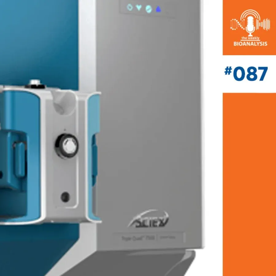
In episode 87 of The Weekly Bioanalysis podcast, John Perkins and Dawn Dufield preview the upcoming WRIB conference, where KCAS Bio and Sciex will both have strong representation through scientific presentations, panels, and business development efforts. Special guest, Rahul Baghla of Sciex, discusses the company’s collaboration with KCAS Bio and introduces their…
 Blogs
Blogs
The Society of Toxicology’s SOT 2025 meeting in Orlando, Florida, held from March 20-22, was a unique gathering that highlighted both the ongoing challenges and the evolving opportunities within the toxicology and bioanalytical fields. As a Bioanalytical CRO providing critical dose formulation testing and toxicology studies…
 News
News
Press Release • Mar 26, 2025 PALO ALTO, Calif., March 26, 2025 (Newswire.com) – Bioz, Inc., the leader in citation management, is highlighting its partnership with KCAS Bio, a trusted provider of bioanalytical and biomarker services. Through the integration of a Bioz Content Hub, KCAS…
 webinars
As the field of biotherapeutics rapidly evolves, the development of advanced conjugated therapies such as Antibody-Drug Conjugates (ADCs), Antibody-RNA Conjugates (ARCs), siRNA/oligos, and antibody-peptide conjugates has gained significant momentum. These next-generation therapeutics offer promising efficacy and safety profiles for treating various conditions, including cancer, rare diseases, and in vaccine development.
 Blogs
Blogs
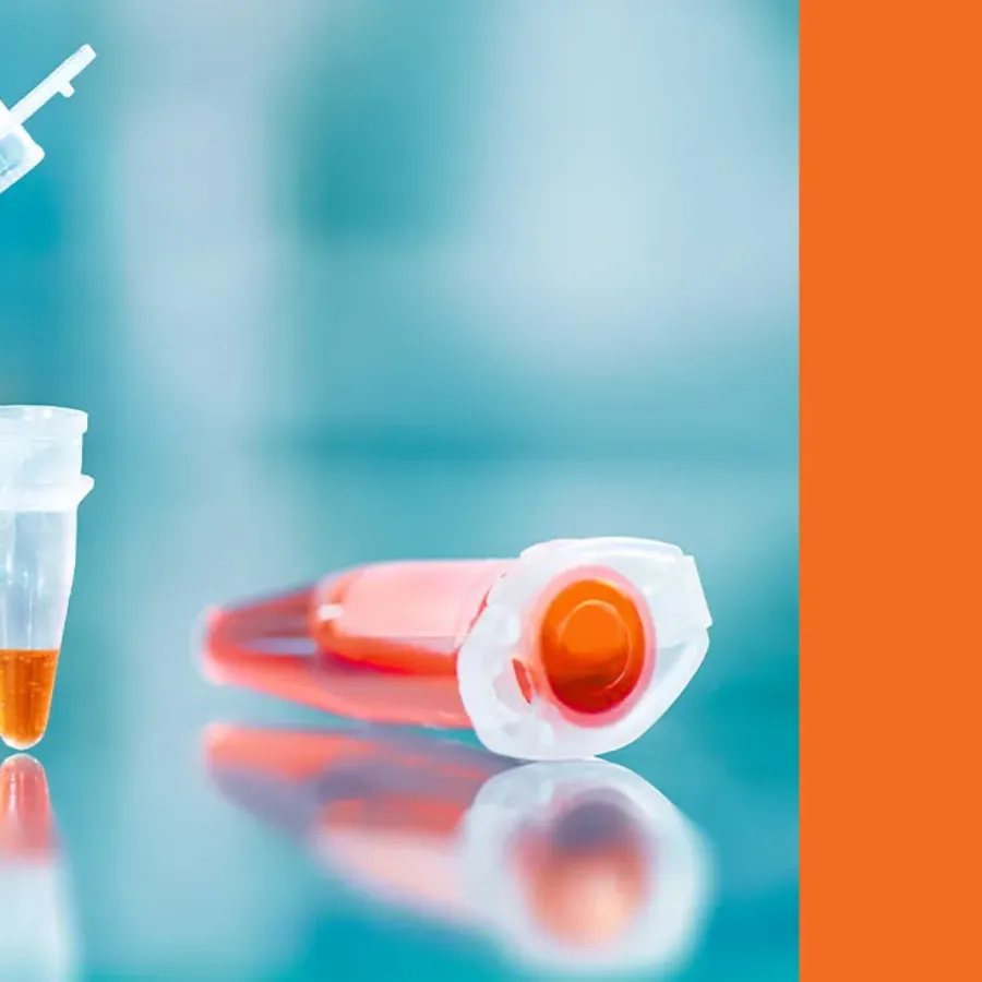
Polymerase Chain Reaction (PCR) has revolutionized molecular biology by enabling the rapid and precise amplification of DNA sequences. Since its invention by Kary Mullis in the 1980s, PCR has become an indispensable tool in both research and diagnostic applications. From identifying genetic disorders to detecting infectious diseases, PCR’s versatility has…
 Blogs
Blogs
KCAS Bio is excited to announce its participation in the upcoming 19th Workshop on Recent Issues in Bioanalysis (WRIB), taking place April 7-11, 2025, in New Orleans, Louisiana. As a long-standing attendee and active contributor at WRIB, KCAS Bio is proud to continue its tradition of…
 Podcasts
In this engaging episode of the podcast, hosts Brian Wile and Adam Cotty welcome special guests Michael Naso, Hillary Quinn, and Barry Morse from The Therapeutic Innovations Group—a powerhouse team of scientists and biotech veterans with decades of experience across biologics, small molecules, and cell therapy. The conversation dives deep…
 Blogs
Blogs
Last month, Elodie, Magali, and I found ourselves in Lisbon—not for a spring getaway (though the weather was a delightful preview of the season) and certainly not for ultra-trail training (although the city’s hills did give our calves a solid workout!). Instead, we were there for EIP, an event…
 Blogs
Blogs
The bioanalysis world has exploded with the need for molecular assays (qPCR, dPCR, NGS, Hybridization technologies) due to the demand for both biodistribution/PK and PD/BM analysis of various drug modalities. Many of these molecular assays have been around for decades and are now routine methods. CLIA and reference labs have…
 Blogs
Blogs
When it comes to developing new therapies for inflammatory bowel diseases (IBD), having robust and reliable bioanalytical support is crucial. From the early stages of drug discovery to Phase III clinical trials, our comprehensive bioanalytical solutions are designed to enhance and accelerate your drug development journey in a GCP…