 Blogs
Blogs
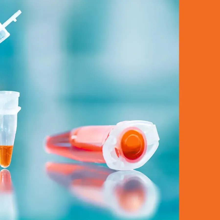
Polymerase Chain Reaction (PCR) has revolutionized molecular biology by enabling the rapid and precise amplification of DNA sequences. Since its invention by Kary Mullis in the 1980s, PCR has become an indispensable tool in both research and diagnostic applications. From identifying genetic disorders to detecting infectious diseases, PCR’s versatility…
 Blogs
Blogs
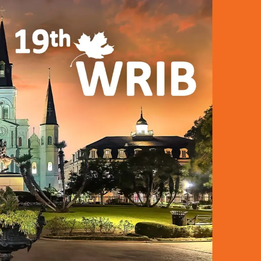
KCAS Bio is excited to announce its participation in the upcoming 19th Workshop on Recent Issues in Bioanalysis (WRIB), taking place April 7-11, 2025, in New Orleans, Louisiana. As a long-standing attendee and active contributor at WRIB, KCAS Bio is proud to continue its tradition…
 Podcasts
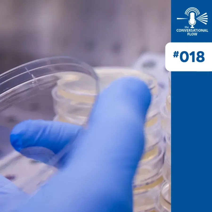
In this engaging episode of the podcast, hosts Brian Wile and Adam Cotty welcome special guests Michael Naso, Hillary Quinn, and Barry Morse from The Therapeutic Innovations Group—a powerhouse team of scientists and biotech veterans with decades of experience across biologics, small molecules, and cell therapy. The conversation dives…
 Blogs
Blogs
Last month, Elodie, Magali, and I found ourselves in Lisbon—not for a spring getaway (though the weather was a delightful preview of the season) and certainly not for ultra-trail training (although the city’s hills did give our calves a solid workout!). Instead, we were there for EIP, an…
 Blogs
Blogs
The bioanalysis world has exploded with the need for molecular assays (qPCR, dPCR, NGS, Hybridization technologies) due to the demand for both biodistribution/PK and PD/BM analysis of various drug modalities. Many of these molecular assays have been around for decades and are now routine methods. CLIA and reference labs…
 Blogs
Blogs
When it comes to developing new therapies for inflammatory bowel diseases (IBD), having robust and reliable bioanalytical support is crucial. From the early stages of drug discovery to Phase III clinical trials, our comprehensive bioanalytical solutions are designed to enhance and accelerate your drug development journey in a…
 Podcasts
During episode 86 of KCAS Bio’s “The Weekly Bioanalysis” podcast, our hosts Dom and John are joined by Dr Cheikh Kane, the Vice President of Biopharma Services at KCAS Bio, to discuss immunogenicity. They first define Immunogenicity; what it is and why it is tested, as well as the…
 Blogs
Blogs
ADCs represent a promising class of targeted therapies, and understanding the intricacies of their analysis is crucial for their successful development. In this blog, we address common questions to guide you through the various considerations involved in ADC research and testing. We will cover key topics related to…
 Blogs
Blogs
Understanding the intricate relationship between drugs and the human body is crucial for effective medical treatments. Two fundamental concepts in pharmacology – pharmacokinetics and pharmacodynamics – play pivotal roles in this understanding. This blog post will delve into these concepts, exploring their differences and significance in drug development and…
 Blogs
Blogs
Cell-based immunotherapies like Chimeric Antigen Receptor (CAR)-T cell therapy have been transforming the treatment of hematologic malignancies for more than a decade. Several groundbreaking studies (1) have led to a growing interest in applying these cell-based therapies to other T and B cell-mediated diseases. One emerging application of CARs…
 Podcasts
Episode 17 of The Conversational Flow podcast, hosted by Brian Wile and Adam Cotty, dives into the evolving landscape of flow cytometry networking and regulatory guidance, where they analyze recent FDA biomarker validation guidance, comparing it to existing industry standards like CLSI H62. Our hosts explore the challenges of harmonizing flow…
 Blogs
Blogs
If you have trained in the field of immunology, it may not be uncommon to emerge with a self-characterization as either a “B-cell immunologist” or a “T-cell immunologist.” Historically, this distinction arose from years of specialization and the degrees of separation between T-cell and B-cell biology research. Approaches to…