 Blogs
Blogs
Spectral flow has come a long way in recent years, transforming from a mere whisper of possibility to an essential tool in bioanalysis. Initially gaining traction in academic institutions, it quickly caught the attention…
 Blogs
Blogs
Biologics are drugs derived from complex molecules like antibodies. Over the last two decades they have re-emerged as…

 Blogs
Blogs
Business Wire – May 24, 2023 11:00 AM EST – In an effort to continue providing the…
 Blogs
Blogs
Being that it is a relatively “niche” segment of the industry, there are several key areas that need to be considered in the field of molecular services related to cell and gene therapies. First and foremost, it is important to recognize that regulatory guidelines within this field are…
 Blogs
Blogs
Understanding the interactions between drugs and biological systems is critical for the success of a new drug. One key tool in this process is functional assays. Functional assays are customized assays that evaluate the impact of drugs on the functionality of cells. They test for a drug’s specific biological mechanism…
 Blogs
Blogs
Cell and Gene Therapies (CGTs) has an estimated market size value in 2022 of USD 8.22 billion and a revenue forecast in 2030 of USD 24.5 billion. This is a CAGR (compound annual growth rate) of 14.6% from 2022 to 2030. Needless to say, the…
 Blogs
Blogs
Due to its ability to analyze multiple parameters across different cell types within a sample, flow cytometry provides rich and clinically valuable data sets from even small volumes of blood. However, flow cytometry is a challenging platform to master, and requires significant investment in equipment and technical training.
 Blogs
Blogs
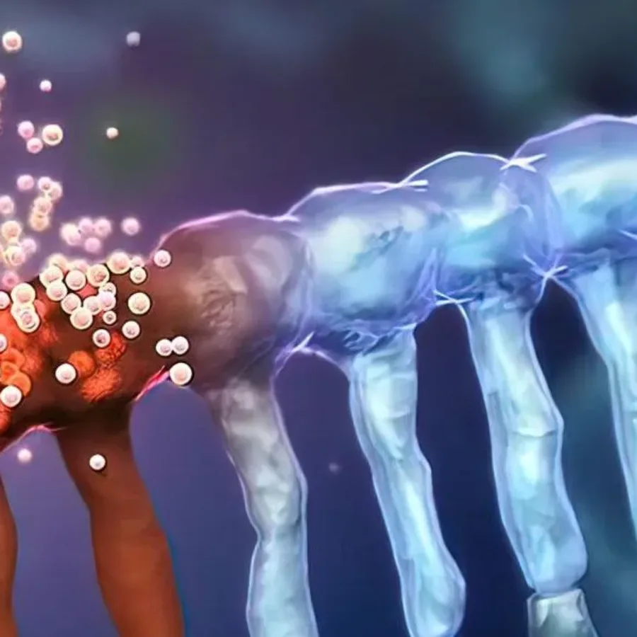
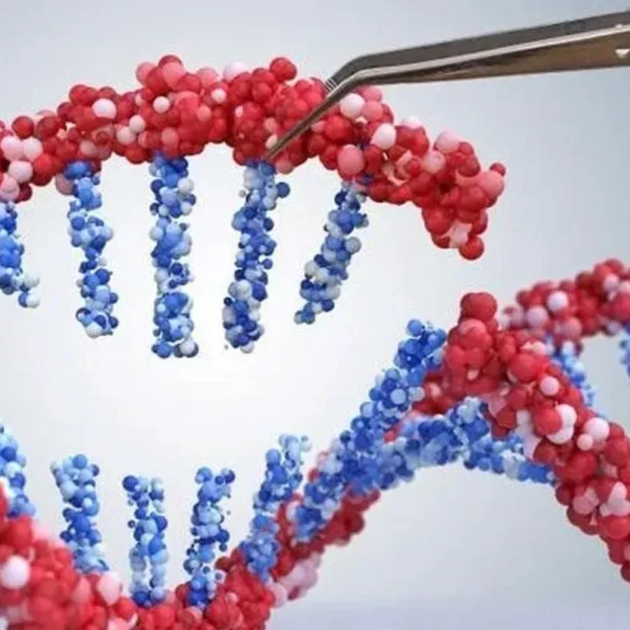
With the extensive advances in technologies like CRISPR and CAR-T, cell and gene therapy has grown to become a viable way for treating Cancer as well as other diseases. Our team has over 100+ years of collective expertise in molecular services using qPCR and ddPCR for support of…
 webinars
Being produced by Xtalks on Friday, June 24, 2022 | 12pm EDT (NA) / 5pm BST (UK) / 6pm CEST (EU-Central) 60 min Webinar Description: Cell and gene therapies (CGTs) are types of treatment that use cellular or genetic material with the goal of treating a disease or a…
 Blogs
Blogs
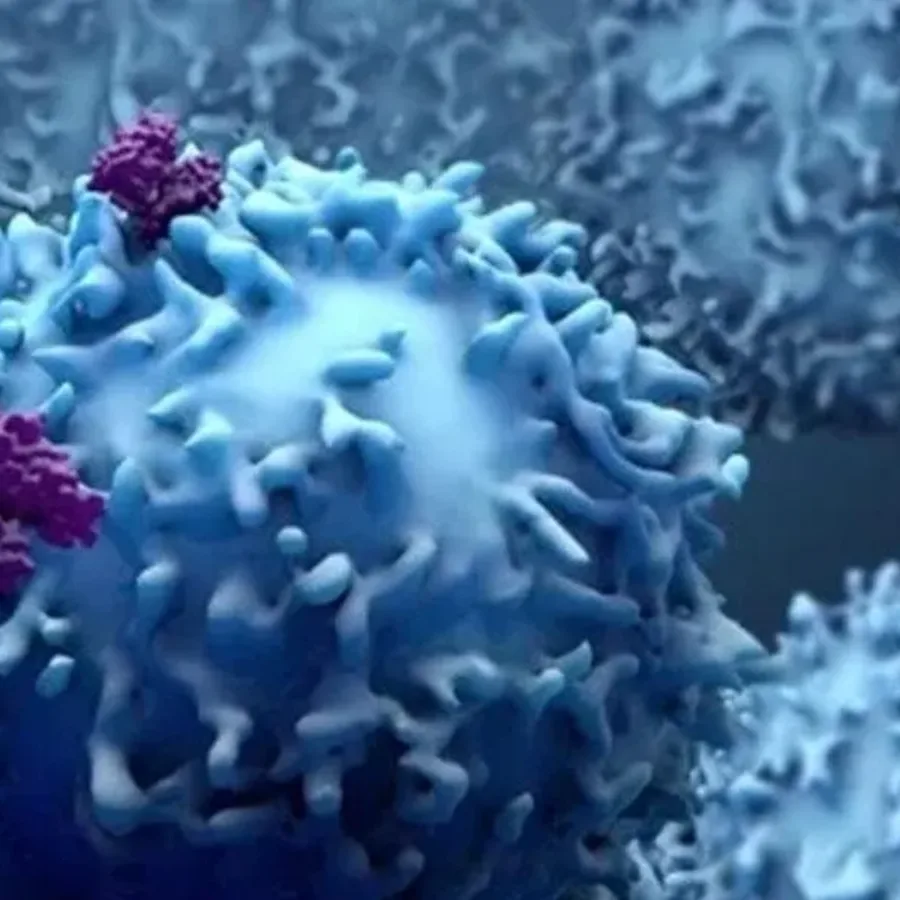
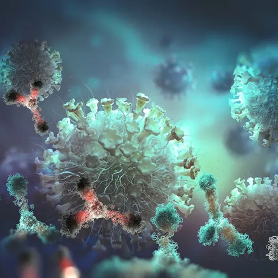
Immunotherapy research is a rapidly expanding field with a pipeline of monoclonal antibodies in development to treat a range of cancers and autoimmune diseases. The mechanism of action (MOA) used by an antibody to mediate a therapeutic response must be fully defined to enable a candidate antibody to advance down the preclinical development pipeline. It is also required for all antibodies used in clinical research and regulatory IND filings in order to optimize dosing and assess the risk of detrimental side effects.
Drugs and biologics in the research pipeline must undergo stringent preclinical toxicology and safety assessments before use in a clinical trial. Most traditional preclinical toxicology screenings include testing in animal models in order to define toxicological and pharmacological parameters that are critical to determining appropriate dosing such as maximum tolerated dose (MTD).