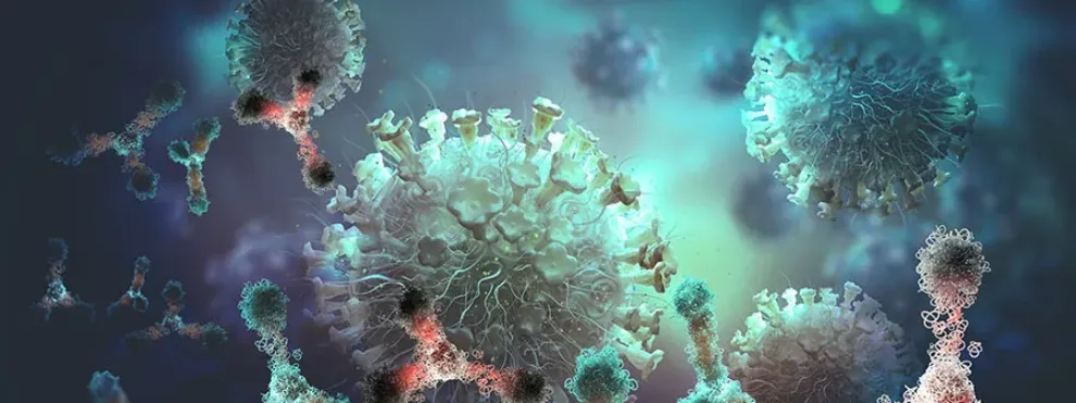
Immunotherapy research is a rapidly expanding field with a pipeline of monoclonal antibodies in development to treat a range of cancers and autoimmune diseases. The mechanism of action (MOA) used by an antibody to mediate a therapeutic response must be fully defined to enable a candidate antibody to advance down the preclinical development pipeline. It is also required for all antibodies used in clinical research and regulatory IND filings in order to optimize dosing and assess the risk of detrimental side effects.
Cytometry to Determine Mechanisms of Action for Therapeutic Antibodies
Therapeutic antibodies have been shown to primarily function through one of six major MOAs to mediate their therapeutic effect; these include blockade, apoptosis, antibody-dependent cellular phagocytosis (ADCP), antibody-dependent cellular cytotoxicity (ADCC), complement-dependent cytotoxicity (CDC) and trogocytosis. Flow cytometry represents the only cellular analysis platform that can assess all six of these MOAs for an experimental antibody. These types of flow cytometry assays are compatible with the assessment of almost all antibodies / antibody-derivatives and cell types and are therefore ideal for translational, preclinical, and clinical studies (Miller, M and Finn, O. 2020).
Blockade Assays
The blockade is a frequently observed mechanism by which a therapeutic antibody interferes with the binding of a target cell receptor or surface molecule and therefore blocks downstream immunological responses (e.g., immune checkpoint inhibitors). Blockade can be monitored using different flow cytometry-based approaches such as receptor occupancy assays. There are various forms of RO assay formats, and these can be used to assess the MOA of a therapeutic antibody.

Apoptosis Assays
Flow Cytometry-based assays for apoptosis can be used to track and quantify the effect of an experimental antibody on target cells within a heterogeneous population through a variety of different biomarkers. Apoptosis (derived from the Greek word ‘apo plus ptosis’ meaning ‘falling off’) is a process of events characterized by initial cell shrinkage, increased cell permeability through membrane disruption, chromatin condensation leading to DNA fragmentation, and finally cell blebbing and the removal of the apoptotic cells by phagocytosis. These multiple phases can be tracked using various flow cytometry methods, that enable researchers to examine different cellular changes.
The induction of apoptosis is correlated with the expression of several cell surface proteins referred to as death receptors (DRs)- these include CD95 (FAS), CD261, CD262 (DR5/TRAIL-R2), CD120a (TNF-R1), CD120b, all of which can be detected with fluorophore-conjugated antibodies using flow cytometry.
The intrinsically mediated apoptosis pathway is typically monitored through the detection of Bcl-2. The extrinsic mediated apoptosis pathway can be monitored by flow cytometry by measuring caspase 3/7, 8, and poly caspase activity using fluorogenic substrates. Correlating with caspase activation, are changes in membrane symmetry and permeability that can be monitored via Annexin V coupled with a cell-impermeable dye such as 7AAD or PI.
There is growing evidence that apoptosis may represent a key step in the development of immunity, and apoptotic cells are thought to be a source of immunological instructions that may influence many different mechanistic pathways of the immune system (Albert 2004).
Antibody-Mediated Cellular Assays
Antibody-dependent cellular phagocytosis (ADCP) can be measured in a variety of ways by flow cytometry but essentially target and effector cells are independently labeled, and the test antibodies are then added. Binding of the antibodies to the target cell via the Fab region, and to the effector cell Fc receptor via their Fc region would trigger ADCP, and the target cell label signal would be detected within the effector cell population.
Similarly, antibody-dependent cellular cytotoxicity (ADCC) assays can measure the death of antibody-coated target cells by cytotoxic mechanisms from effector cells in PBMCs, primarily natural killer (NK) cells.
Both ADCP and ADCC assays are used to support the development of monoclonal antibody-based therapeutics and the assessment of biosimilars within clinical trials. Since IND filing now requires MOA profiling, these two assay types are employed across many areas of medicine including tumor biology and immuno-oncology.
Complement-dependent cytotoxicity (CDC) assays are used to quantify the death of target cells bound with antibodies that interact with components of the complement system and activate the complement cascade, resulting in target cell lysis. CDC assays are frequently employed to determine the efficacy of antibody-based therapeutics and their capacity to initiate multiple facets of the immune system to kill targeted cells (Gancz, D. and Fishelson, Z, 2009), as well as assess xeno-transplants.

Trogocytosis Assays
Trogocytosis (derived from the ancient Greek ‘trogo’, meaning ‘gnaw’) is a mechanism by which lymphocytes (B, T, or NK cells) capture fragments of plasma membrane from the antigen-presenting cells that are expressing their cognate antigen. Essentially, this process enables the extraction of the antibody-ligand complexes from the target cell membrane and subsequent transfer of the complexes to the lymphocyte membrane. The transfer of the antibody complex from target cell to lymphocyte can be measured by flow cytometry by pre-labelling the target cell plasma membrane with a lipophilic dye such as PKH67B; following co-incubation of the labelled APCs and lymphocytes, trogocytosis is monitored through the increased staining of the lymphocyte cells with this label over time. Trogocytosis assays are being used to study the tumor microenvironment and the development of chemoresistance (Rafli, A. et. al. 2008). It is thought that trogocytosis by cytotoxic T-cells may enhance the selection of higher-affinity T-cells through the harvesting of antigenic complexes from antigen-presenting dendritic cells (Kedl et. al. 2002).
Final Thoughts
The development of therapeutic monoclonal antibodies represents one of the most active areas of drug research and development, with the potential to be used to treat patients with cancer, autoimmune- and infectious diseases through the modulation of the immune system. These biological drugs can function through multiple different mechanisms, and it is essential to understand which are the driving responses to select the most active and least toxic monoclonal antibodies for targeted clinical applications. The six MOAs outlined here trigger many different types of downstream immune responses depending on the target and effector cells involved. Functional MOA assays can be used to select for desired characteristics as well as support mutational screening for increased antibody potency or reduced toxicity. Flow cytometry-based MOAs represents a high throughput platform that enables these complex biological drug candidates to be more effectively analyzed, across a wide scope of the immune responses.
References
- Miller, M. and Finn, O. Chapter Twenty-Two – Flow cytometry-based assessment of direct-targeting anti-cancer antibody immune effector functions, Editor(s): Lorenzo Galluzzi, Nils-Petter Rudqvist, Methods in Enzymology, Vol. 632, 2020, Pp. 431-456, ISSN 0076-6879, ISBN 9780128186756, https://doi.org/10.1016/bs.mie.2019.07.026
- Albert, M. Death-defying immunity: do apoptotic cells influence antigen processing and presentation? Nat. Rev Immunol 4, 223–231 (2004). https://doi.org/10.1038/nri11308
- Dana Gancz, Zvi Fishelson. Cancer resistance to complement-dependent cytotoxicity (CDC): Problem-oriented research and development. Molecular Immunology, Volume 46, Issue 14, 2009, Pp. 2794-2800, ISSN 0161-5890, https://doi.org/10.1016/j.molimm.2009.05.009
- Rafii A, Mirshahi P, Poupot M, Faussat A-M, Simon A, et al. (2008) Oncologic Trogocytosis of an Original Stromal Cells Induces Chemoresistance of Ovarian Tumours. PLoS ONE 3(12): e3894. doi:10.1371/journal.pone.0003894
- Kedl, R.M. et. al. T cells down-modulate peptide-MHC complexes on APCs in vivo. Nat. Immunol. 3, 27–32 (2002)

