 November 15
- November 17
November 15
- November 17
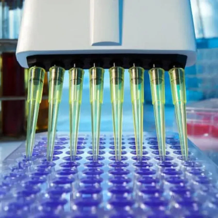
For years, Dawn Dufield, PhD and I have been doing our best to get the word out (via as many venues as possible) about KCAS’s unique expertise with hybrid LC-MS/MS. This year’s upcoming EBF meeting in Barcelona is just such an opportunity, and one we are very much looking…
 Blogs
Blogs
How Do Drug Indications Change During Development? The Pharm/Biotech industry’s main focus is on development of therapeutics to address unmet medical needs but sometimes, clinical trials lead to observations that lead to a change in research path for a drug. A good example is sildenafil that was originally studied for…
 Blogs
Blogs
In the ever-evolving landscape of pharmaceutical research, biodistribution studies play a pivotal role in understanding how drugs and therapies are distributed within the body. These studies are critical for assessing the efficacy and safety of new treatments, particularly in the realm of cell…
 Blogs
Blogs
Recently, Matthew Pennington, PhD (one of KCAS’s Senior Scientists) was the senior author on a publication entitled “A Validated Droplet Digital Polymerase Chain Reaction Assay for the Detection of Adeno Associated Viral Vectors in Bio Shedding Studies of Tears”. Below, Matt explains the significance in what they accomplished…
 Blogs
Blogs
In the rapidly evolving realm of flow cytometry, conventional flow cytometry is gradually being displaced by its advanced counterpart, spectral flow cytometry. These technologies, each with its unique strengths and applications, have revolutionized the way we analyze cells and particles. Let’s dive…
 Blogs
Blogs
A biomarker is a defined characteristic that is measured as an indicator of normal biological processes, pathogenic processes, or biological responses to an exposure or intervention, including therapeutic interventions(1). With the development of innovative and personalized therapies, biomarker tracking is increasingly important to support the clinical development…
Blogs
Immunophenotyping has undergone a seismic change in less than two decades as panel sizes have increased in complexity from <10 to >40 colors. Let’s explore how immunophenotyping is transforming the field and how KCAS Bio is at the forefront of this…
 Blogs
Blogs
In an exciting development, KCAS, through its subsidiary FlowMetric, is expanding its flow services in Europe. With a history of providing cutting-edge flow services in the EU, KCAS is taking a significant step forward by transitioning services from its Milan, Italy site to…
 Blogs
Blogs
With recent guidance released from the FDA, there are changes for PKs (Pharmacokinetics) and ADCs (Antibody Drug Conjugates) that must be clearly understood before making decisions for your drug product testing. ADCs combine the target specificity of monoclonal antibodies with the…
 Blogs
Blogs
Spectral flow has come a long way in recent years, transforming from a mere whisper of possibility to an essential tool in bioanalysis. Initially gaining traction in academic institutions, it quickly caught the attention…
 Blogs
Blogs
In the ever-evolving landscape of pharmaceutical research…
 Blogs
Blogs
Biologics are drugs derived from complex molecules like antibodies. Over the last two decades they have re-emerged as…