Posts by KCAS Bio
 Blogs
Blogs
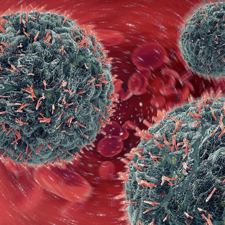
Immunotherapeutic molecules currently being used in the clinic are powerful immune modulators, but their effectiveness can be inconsistent between patients. Clinicians and scientists use different assays to evaluate why immunotherapies fail in the clinic. The flow cytometry-based receptor occupancy (RO) assay is a critical tool for evaluating the effectiveness of immunotherapies in the clinic. Here are three features of flow cytometry-based RO assays that give them clinical value.
 Blogs
Blogs
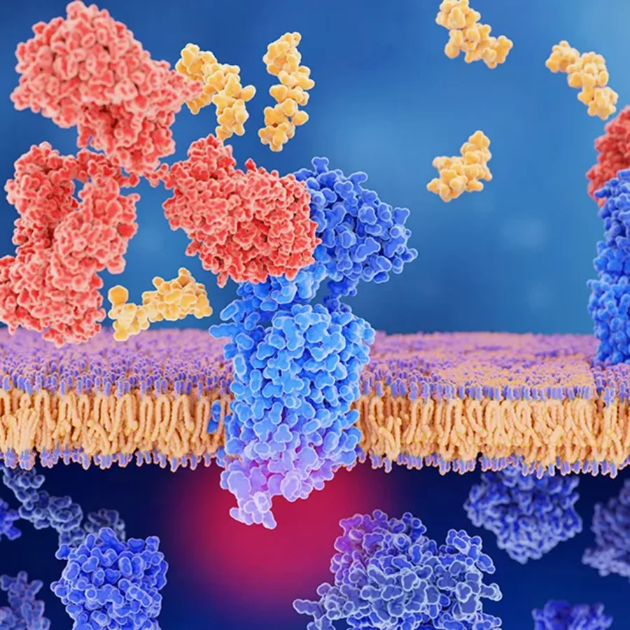
What are receptor occupancy assays?
 Blogs
Blogs
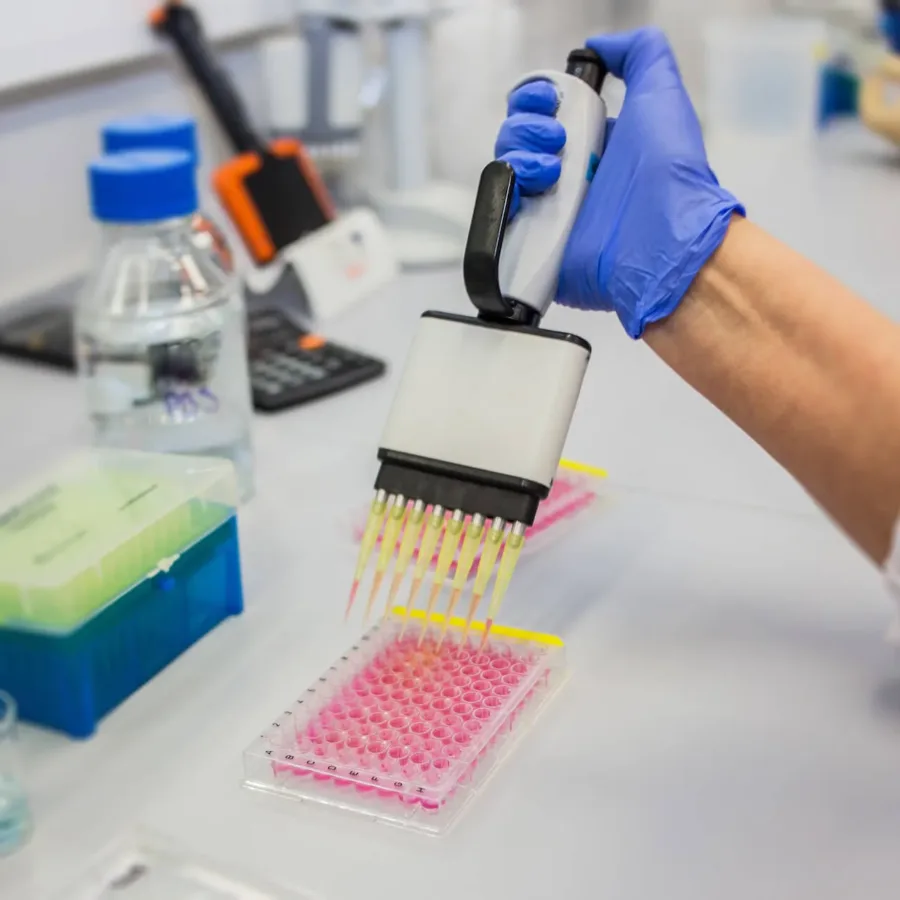
Q is for Quality - QA, QC and Flow Cytometry How do clinical flow cytometry labs ensure that the data they generate is accurate, reproducible, and conforms to regulatory requirements? They use quality management systems, including quality assurance (QA) and quality control (QC). Some scientists seem to use these terms interchangeably, but what do they really mean and why are they important to flow cytometry?
 Blogs
Blogs
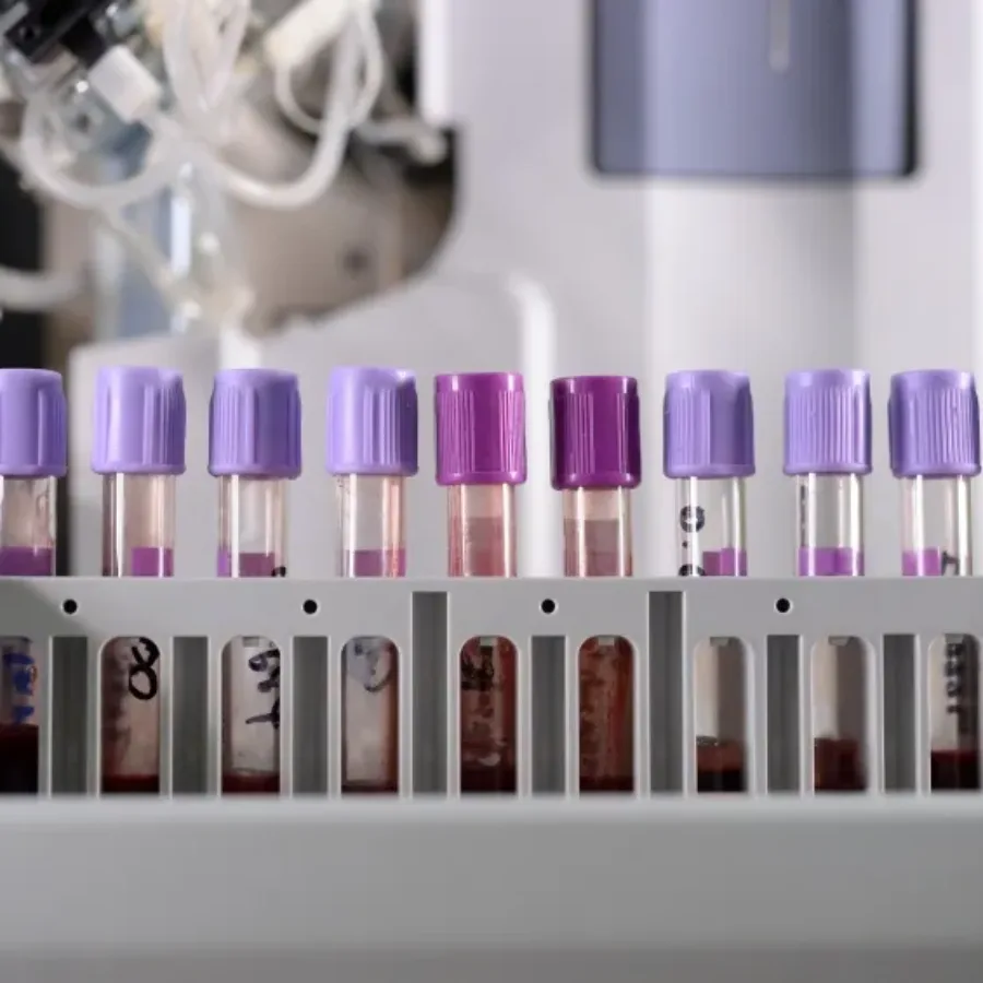
Introduction of CAR-T Therapy T lymphocytes are engineered with synthetic receptors known as chimeric antigen receptors (CAR) in CAR-T Cell therapy. The CAR-T cell is an effector T cell that recognizes and eliminates specific cancer cells, independent of major histocompatibility complex molecules. (Zhai et al. 2018). Chimeric antigen receptors (CARs) cells have recombinant receptor constructs expressed in T cells to target cells expressing specific antigens.
 Blogs
Blogs
Flow cytometry is a powerful tool for surveying the cellular landscape during preclinical development of drugs and biologics. But flow cytometry can go beyond immunophenotyping to actual functional measurements that can contribute to understanding the true potential of a therapeutic candidate. To make the most of your flow cytometry studies, consider these other assays as you plan the next phase of preclinical development.
 Blogs
Blogs
Flow cytometry is a powerful tool because it allows users to analyze the characteristics of millions of cells with relative speed and precision. A single cell suspension of fluorescently labeled sample travels through the cytometer for excitation by lasers, and the emitted light photons are measured by different detectors. Having a single cell suspension is essential to measuring cell fluorescence accurately, and many types of cell or tissue samples must be specially processed to make this suspension. Two different methods can be used for single cell suspensions: mechanical dissociation of tissue or enzymatic dissociation of tissue. This processing step is typically carried out before cells are stained and both methods have benefits and caveats.
 Blogs
Blogs
Flow Cytometry utilizes fluorescently labeled antibodies to detect specific biomarkers on the surface and within cells, and over the past few years, there has been a surge in reagents available for flow cytometry applications. Most of these have been developed using monoclonal antibodies raised in mice and conjugated to a range of fluorophores. However, there are still instances where suitable monoclonal antibody reagents/conjugates are not commercially available, and small-scale conjugations are not practical. In these instances, so-called indirect staining may be employed, where the binding of an unconjugated primary antibody is detected using a secondary anti-IgG antibody conjugate.
 Blogs
Blogs
Memory is a characteristic of the immune system that provides humans and other vertebrates with long term protection against infectious diseases and other “non-self” antigens such as those associated with tumor cells. In the context of T cells, memory responses occur when a naïve T cell encounters an antigen bound to a major histocompatibility complex molecule and is activated to undergo differentiation into an effector cell or a memory cell. Memory T cell populations can persist in the body for months to years and can be stimulated to respond specifically and rapidly to a foreign antigen upon re-exposure.
 Blogs
Blogs
Flow cytometry is a powerful technique for characterizing immune responses to vaccines, immunotherapeutic drugs, and other clinical interventions. But many preclinical and clinical studies may take place at sites that are not in the same location as the flow cytometry lab. That’s why it’s critical to determine how clinical specimens should be collected, processed, stored, and shipped to assure that cells will be viable and abundant enough for flow cytometry analysis.
 Blogs
Blogs
The flow cytometry market is filled with an abundance of products for mouse and human samples. But what if your studies use different species? Fortunately, many antibodies for standard cell markers can work on multiple species, and more species-specific reagents are becoming available.
 Blogs
Blogs
With the rapid progress of immune-monitoring drug development, flow cytometry has found itself increasingly at the forefront of clinical trial assessment of safety and efficacy. This is not without challenges since flow cytometry analysis can be complicated and expensive, too often employs idiosyncratic experimental and analytical methods. So how can a platform without standardized methods and processes, be successfully applied to evaluate clinical endpoints?
