 Blogs
Blogs
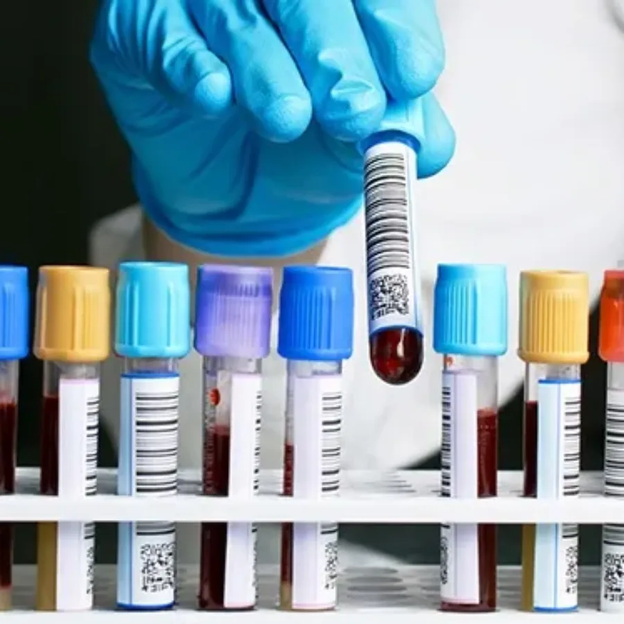
Assay validation is required during the development of new drugs or biologics in order to be in compliance with regulatory requirements for all studies that are not considered research/exploratory in nature. Beyond compliance, what is the point of assay validation? Assays must be precise, robust, and specific during use in preclinical studies and clinical trials in order to assure that drug candidates can be accurately evaluated for safety and efficacy. Validation plans assure that an assay will work reliably, even if an assay is run at different sites or by different users. Consider these other elements of assay validation to understand why it is a critical to preclinical and clinical research: Fit-for-purpose strategy: Each validation plan determines if an assay is made to be fit-for-purpose for a given client’s evaluation needs. Assays can be customized to meet the specific needs of a preclinical screening or clinical trial and fit-for-purpose validation assures that reliable data can be obtained from assay. Test scripts: Test scripts are a series of procedures to be executed during a validation in order to determine if an assay satisfies the necessary specifications or to reveal errors that must be addressed. Running test scripts is essential to the development and continued reliability of a validated assay. GLP compliance: Assay validation must be carried out under the same conditions as will be used for routine assay use. Good laboratory practices (GLP) conditions are needed for many preclinical and clinical applications, and routine quality assurance/quality control monitoring may also be necessary. Be aware of these special circumstances during assay validation. Validation is so much more than a regulatory hurdle. Validation gives you confidence that an assay will yield reliable results that can be trusted to make critical decisions for advancing drug candidates or evaluating clinical efficacy.
 Blogs
Blogs
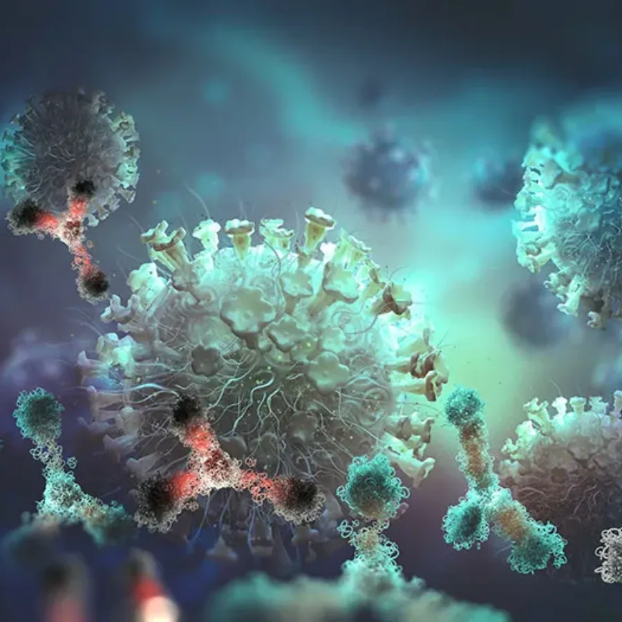
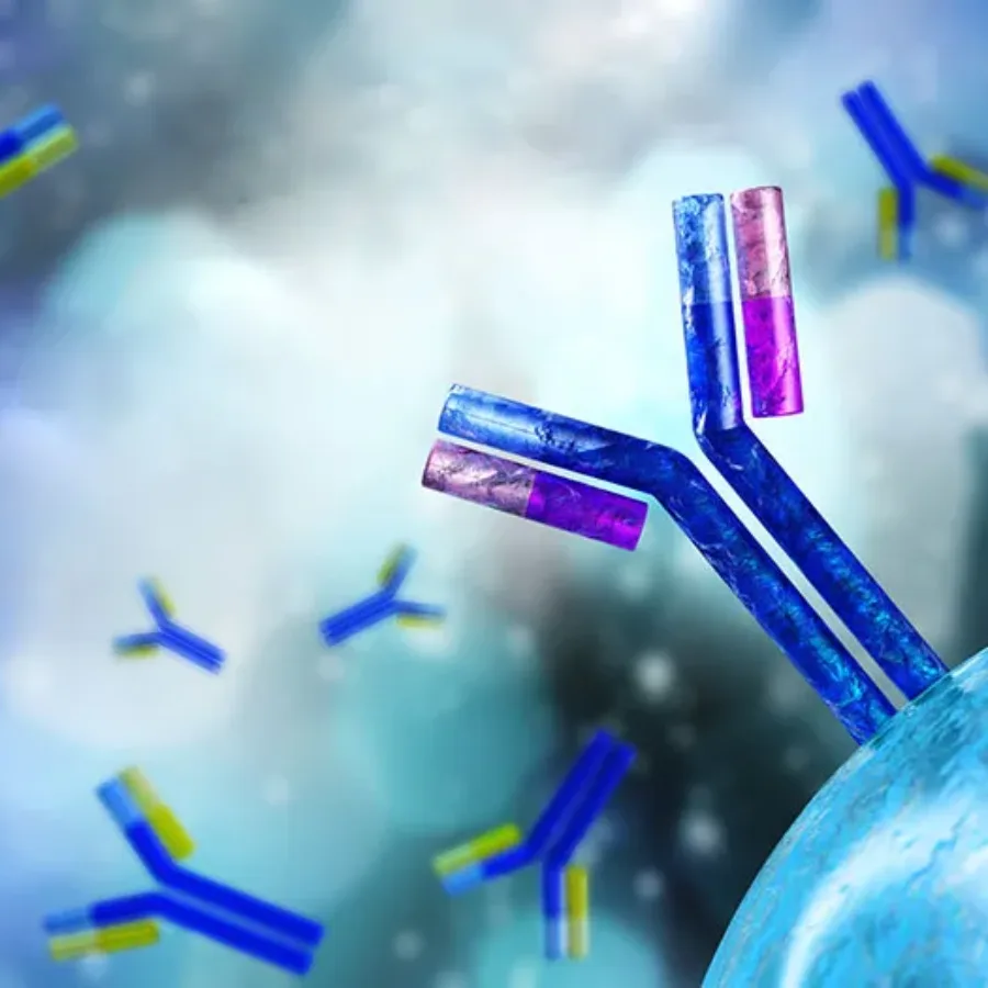
Immunotherapy research is a rapidly expanding field with a pipeline of monoclonal antibodies in development to treat a range of cancers and autoimmune diseases. The mechanism of action (MOA) used by an antibody to mediate a therapeutic response must be fully defined to enable a candidate antibody to advance down the preclinical development pipeline. It is also required for all antibodies used in clinical research and regulatory IND filings in order to optimize dosing and assess the risk of detrimental side effects.
 Blogs
Blogs
Flow cytometry has been developed and used as a clinical tool since the invention of the first cytometers in the 1970s. At present, flow cytometry is considered essential for many routine clinical diagnostics, including assays for leukemia and lymphoma, stem cell enumeration, solid organ transplantation, HIV infection status, immunodeficiencies, and hematologic abnormalities. Many scientists involved in clinical trials or drug development are faced with developing clinical flow cytometry assays for multiple phases of clinical development. If you find yourself starting to plan a clinical flow cytometry assay, here are the top 3 issues to think about as you plan your experiment.
 Blogs
Blogs
Flow cytometry is an elegant and powerful tool that has been critical to understanding the immune system and advancing the development of immune-based therapies. Critical to many studies, and essential for FDA filings, is the development and documentation of a validated assay. While most flow cytometric assays fall into the “quasi-quantitative” category according to FDA guidelines, there are some assays that can be quantitative and even qualitative.
 Blogs
Blogs
Flow Cytometry utilizes fluorescently labeled antibodies to detect specific biomarkers on the surface and within cells, and over the past few years, there has been a surge in reagents available for flow cytometry applications. Most of these have been developed using monoclonal antibodies raised in mice and conjugated to a range of fluorophores. However, there are still instances where suitable monoclonal antibody reagents/conjugates are not commercially available, and small-scale conjugations are not practical. In these instances, so-called indirect staining may be employed, where the binding of an unconjugated primary antibody is detected using a secondary anti-IgG antibody conjugate.

 Blogs
Blogs
Receptor occupancy (RO) assays are a powerful tool for pharmacokinetic/pharmacodynamic evaluations of candidate drugs and biologics. RO assays can also be used toward dose selection for candidate molecules being evaluated in clinical trials. Flow cytometry-based RO assays are currently being used in many sectors of biopharmaceutical drug development. Consider these five things to know about RO assays if you are planning to use this type of assay in your preclinical research.
 Blogs
Blogs
Phosphoflow cytometry assays are becoming a valuable tool for researchers developing immuno-oncology applications because data from these assays can provide critical mechanistic insights. Phosphoflow assays measure phosphorylated proteins in cells, which is a critical readout for cell signaling responses. Check out these five facts about phosphoflow cytometry and consider adding this tool to your cytometry toolbox.
 Blogs
Blogs
With the rapid progress of immune-modulating drug development, flow cytometry has found itself increasingly at the forefront of clinical trial assessment of safety and efficacy. This is not without challenges since flow cytometry analysis can be complicated and expensive, too often employs idiosyncratic experimental and analytical methods. So how can a platform without standardized methods and processes, be successfully applied to evaluate clinical endpoints?
 Blogs
Blogs
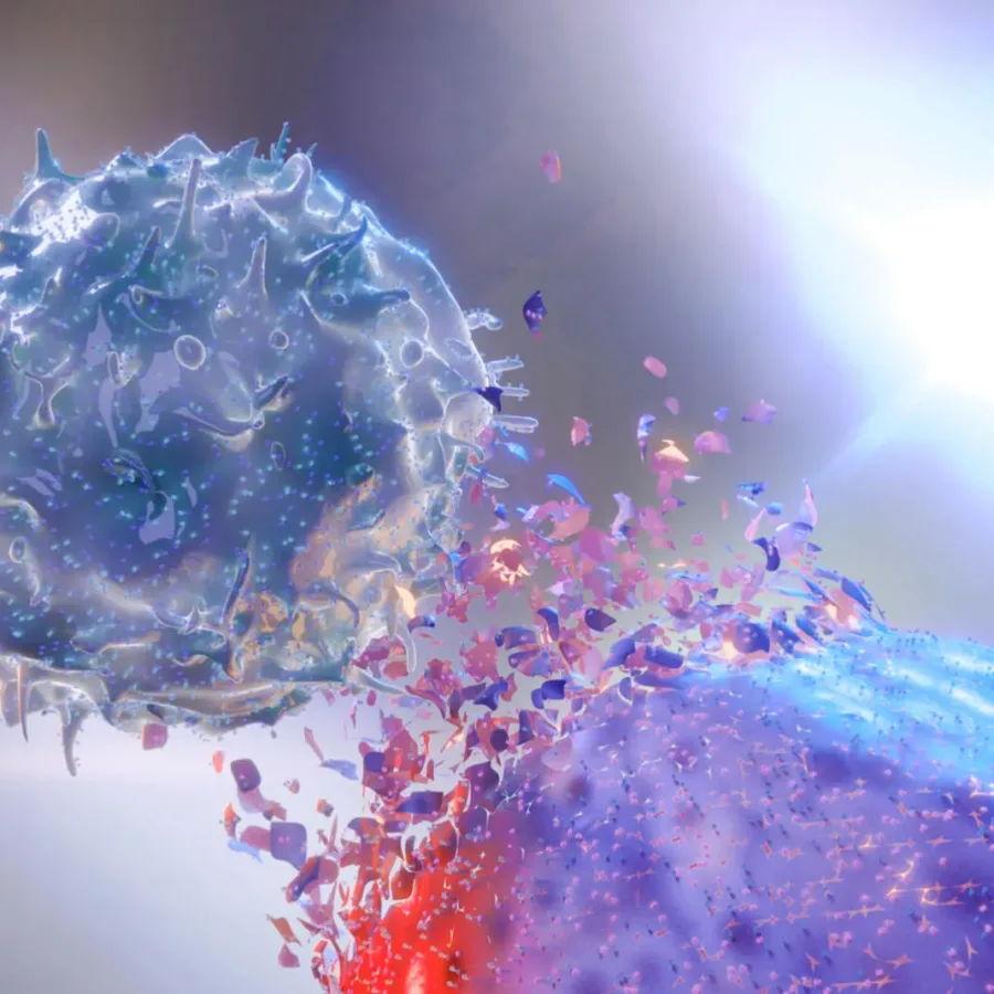
What is the primary role of Natural Killer (NK) cells? Natural killer (NK) cells are the predominant innate immune cells that mediate anti-tumor and anti-viral responses, and therefore possess good clinical utilization (Abel et al. 2018). Natural killer cells comprise 10–15% of peripheral blood lymphocytes and classically display a half-life of approximately 7–10 days in the circulation (Moretta et al. 2000).
 Blogs
Blogs
Although bioanalysis is KCAS’ principal area of expertise, we are always considering how we can build around those capabilities to ensure that we can provide optimal service for our clients. Our current breadth of service enables us to provide a comprehensive approach during non-GLP pre-clinical studies where it is feasible…
 Blogs
Blogs
As a Senior Director for Discovery in the pharma division at KCAS, I would like people to think of “Bioanalytical Discovery” as a stage in drug development; one where you are doing your final…