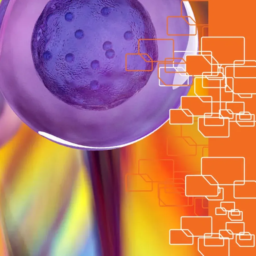
therapeutic_antibodies_featured Blogs
Read article Flow Cytometry for Therapeutic Antibody Assessment

Immunotherapy research is a rapidly expanding field with a pipeline of monoclonal antibodies in development to treat a range of cancers and autoimmune diseases. The mechanism of action (MOA) used by an antibody to mediate a therapeutic response must be fully defined to enable a candidate antibody to advance down the preclinical development pipeline. It is also required for all antibodies used in clinical research and regulatory IND filings in order to optimize dosing and assess the risk of detrimental side effects.

Blogs
Read article Flow Cytometry in Preclinical Toxicology

Drugs and biologics in the research pipeline must undergo stringent preclinical toxicology and safety assessments before use in a clinical trial. Most traditional preclinical toxicology screenings include testing in animal models in order to define toxicological and pharmacological parameters that are critical to determining appropriate dosing such as maximum tolerated dose (MTD).

biology research scientist working in lab Blogs
Read article Using Animal Samples in Flow Cytometry for Toxicology Studies

Toxicology screening is essential to any preclinical study, and flow cytometry-based toxicology assays are a fast and practical approach. Different animal models are used for toxicology studies, including rodents, dogs and non-human primates. One commonly used technique is the micronucleated erythrocyte endpoint assay, which measures DNA damage induced by exposure to experimental drugs or biologics. This method has been adapted into a validated flow cytometry-based assay and can use erythrocytes from different species. Consider these factors when selecting the best toxicology screening method.

alpha_soup Blogs
Read article Developing Clinical Flow Cytometry Assays

Flow cytometry has been developed and used as a clinical tool since the invention of the first cytometers in the 1970s. At present, flow cytometry is considered essential for many routine clinical diagnostics, including assays for leukemia and lymphoma, stem cell enumeration, solid organ transplantation, HIV infection status, immunodeficiencies, and hematologic abnormalities. Many scientists involved in clinical trials or drug development are faced with developing clinical flow cytometry assays for multiple phases of clinical development. If you find yourself starting to plan a clinical flow cytometry assay, here are the top 3 issues to think about as you plan your experiment.

iStock-538032878 Blogs
Read article Tips for Measuring T Cell Proliferation by Flow Cytometry

Many basic and clinical immunology studies that focus on T cells include proliferation assays in order to determine if T cells are capable of proliferating under different in vitro or in vivo conditions. Flow cytometry is the ideal approach for measuring T cell proliferation and a suite of staining products…

Diversity-of-AML-and-FLT3-Phosphorylation-Mutations_COVER-IMAGE Blogs
Read article The Diversity of AML and Prominence of Phosphorylation in FLT3+ Therapy Resistance

Anyone who starts an investigation of acute myeloid leukemia (AML) soon finds out the complexity of this disease. Although daunting initially, it soon becomes apparent the need for complex classifications for AML subtypes and different mechanisms for formation. AML forms from a wide variety of DNA mutations leading to numerous phenotypic changes in the blood makeup. Early on there were the French-American-British classifications in the 1970s (FAB) but in present day, AML type is being broken down to genetic markers. For the most part, this is due to the advancement of scientific-technical capability. Conversely, being able to clearly define AML by mechanistic function, allows for clinicians to state, with some certainty, treatment and survival options for their patients.

iStock-894635270_RT Blogs
Read article T Cell Exhaustion – Using Flow Cytometry to Monitor this Immuno-Oncology Impediment

T cells are well known for their roles in combating cancer and infection, but chronic exposure to antigens and inflammation can cause T cells to enter a state of “exhaustion[1].” Exhausted T cells lose critical effector functions including cytokine production, the ability to proliferate and memory T cell differentiation is also compromised. Exhausted T cells also express inhibitory receptors and become unresponsive to IL-7 and/or IL-15-driven self-renewal. This progression toward T cell exhaustion results in diminished control of chronic infection or cancer. Exhaustion can occur in both CD4+ and CD8+ T cell populations and the phenotypes of these subsets is somewhat heterogeneous. Nonetheless, T cell exhaustion is reversible and various immuno-oncology interventions have been examined or are currently being evaluated in order to improve outcomes in cancer and chronic infection[2].

PATTERN_BLUE_1320x780_3 Blogs
Read article Assay Validation and Compliance in Flow Cytometry

Flow cytometry is an elegant and powerful tool that has been critical to understanding the immune system and advancing the development of immune-based therapies. Critical to many studies, and essential for FDA filings, is the development and documentation of a validated assay. While most flow cytometric assays fall into the “quasi-quantitative” category according to FDA guidelines, there are some assays that can be quantitative and even qualitative.

Fluorescent stem cells under the microscope Blogs
Read article Sorting out the Details: Three Factors to Consider for Cell Sorting by Flow Cytometry

Fluorescent-Activated Cell Sorting (FACS) is a flow cytometry-based technique in which cells are stained with fluorescently labelled antibodies and sorted based on pre-defined staining parameters that are specific to different cell types. FACS users must consider multiple factors when designing and running a FACS experiment. Consider these three factors as you plan and carry out your next FACS experiment.

iStock-174990222 Blogs
Read article Isolating Rare Cell Phenotypes Through Advanced Cytometry

The immune system is comprised of a multitude of unique cell subsets. Each cell type, from B and T cells, to macrophages, monocytes and dendritic cells, have been phenotypically subdivided into unique subsets as we learn more about the phenotypic signatures that define these cells. Flow cytometry has been the central tool in evaluating and defining cell subsets, and major advances in immunophenotyping have occurred recently as more parameters can be measured during a single run on newer flow cytometers.

dreamstimelarge_25505743 Blogs
Read article Some Stimulating Situations for Flow Cytometry

In a healthy person, immune cells like B cells, T cells, and macrophages are typically surveilling the body for abnormal cells or infectious agents. These cells don’t fire up their inflammatory toolbox unless they recognize one of these foreign entities. The potent inflammatory mediators activated during these responses include cytokines, free radicals, prostaglandins, and clotting factors, which must be tightly regulated to avoid wreaking havoc on healthy tissue. This exquisitely controlled activation of inflammatory molecules means that when you look for them in cells by flow cytometry, they may be very difficult to detect.

3 Blogs
Read article Flow Cytometry Sample Preparation – Best Practices

The idiom, garbage in, garbage out applies to many areas of scientific research, including flow cytometry. Good sample preparation is critical to accurate and sensitive cytometry analysis of cells, wherever their origin.