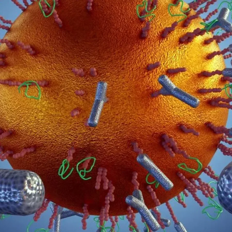
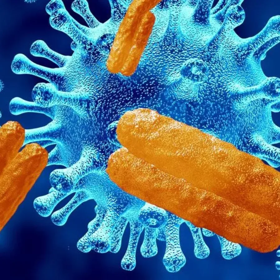
Memory is a characteristic of the immune system that provides humans and other vertebrates with long term protection against infectious diseases and other “non-self” antigens such as those associated with tumor cells. In the context of T cells, memory responses occur when a naïve T cell encounters an antigen bound to a major histocompatibility complex molecule and is activated to undergo differentiation into an effector cell or a memory cell. Memory T cell populations can persist in the body for months to years and can be stimulated to respond specifically and rapidly to a foreign antigen upon re-exposure.
Flow cytometry is a powerful tool because it allows users to analyze the characteristics of millions of cells with relative speed and precision. A single cell suspension of fluorescently labeled sample travels through the cytometer for excitation by lasers, and the emitted light photons are measured by different detectors. Having a single cell suspension is essential to measuring cell fluorescence accurately, and many types of cell or tissue samples must be specially processed to make this suspension. Two different methods can be used for single cell suspensions: mechanical dissociation of tissue or enzymatic dissociation of tissue. This processing step is typically carried out before cells are stained and both methods have benefits and caveats.
Flow cytometry is a powerful technique for characterizing immune responses to vaccines, immunotherapeutic drugs, and other clinical interventions. But many preclinical and clinical studies may take place at sites that are not in the same location as the flow cytometry lab. That’s why it’s critical to determine how clinical specimens should be collected, processed, stored, and shipped to assure that cells will be viable and abundant enough for flow cytometry analysis.
With the rapid progress of immune-monitoring drug development, flow cytometry has found itself increasingly at the forefront of clinical trial assessment of safety and efficacy. This is not without challenges since flow cytometry analysis can be complicated and expensive, too often employs idiosyncratic experimental and analytical methods. So how can a platform without standardized methods and processes, be successfully applied to evaluate clinical endpoints?
The flow cytometry market is filled with an abundance of products for mouse and human samples. But what if your studies use different species? Fortunately, many antibodies for standard cell markers can work on multiple species, and more species-specific reagents are becoming available.
Data by FlowMetric All flow cytometry experiments begin with similar basic set up protocols to assure that the equipment is functioning properly and that samples can be measured accurately. Using a flow cytometry gating strategy is an essential step during this set up phase as it assures that the correct cell populations are being measured. Here are factors to consider as you determine how you want to establish your flow cytometry gating strategy for your next experiment.
Flow cytometry assays are important for preclinical and clinical research, however, it is vital to understand the level of compliance required for the stage of research you are completing. Flow Cytometry assays completed for toxicology and safety assessments are required to be in compliance of Good Laboratory Practices (GLP), on the other hand, basic research or discovery/exploratory studies can be non-GLP. GLP refers to a set of standards for laboratory studies to be planned, performed, monitored, reported, and archived. Preclinical and clinical studies must be GLP-compliant in order to be submitted for review by regulatory agencies like the FDA. Consider these three points if you find yourself in need of a GLP-compliant flow cytometry assay.
There is no question that the discovery of vaccines spearheaded the path of modern medicine and in so doing, eradicated at least two diseases, smallpox, and rinderpest from the global population. Today’s modern vaccines are being developed not only to tackle infectious diseases but also for the treatment and prevention of autoimmune diseases and cancers. Whereas vaccines for infectious diseases and cancer are designed to provoke a specific Th 1-driven immune response to target and reject the tumor or pathogen, vaccines driving Th 2 responses appear to be the best at targeting autoimmune diseases. Understanding the driving factors behind these underlying responses is central to the development of safe and effective vaccines, and flow cytometry provides unprecedented clarity on how the immune system responds to different vaccine strategies.
Chimeric antigen receptor (CAR) T cell therapy is transforming patient-specific cancer treatment, even for the most challenging forms of cancer. CAR T cells are made by isolating a patient’s T cells from the blood and engineering them in the lab so that they can specifically fight the patient’s cancer. This custom-made biologic is both time and labor-intensive and extremely costly, but it is also an extremely effective form of treatment.
Receptor occupancy (RO) assays are a powerful tool for pharmacokinetic/pharmacodynamic evaluations of candidate drugs and biologics. RO assays can also be used toward dose selection for candidate molecules being evaluated in clinical trials. Flow cytometry-based RO assays are currently being used in many sectors of biopharmaceutical drug development. Consider these five things to know about RO assays if you are planning to use this type of assay in your preclinical research.
Fluorescence-activated cell sorting is a powerful tool for basic and clinical research because individual cells can be separated from a heterogeneous sample and used for downstream analysis or therapeutic applications. A fluorescent activated cell sorter works in a similar way as a flow cytometer. A single-cell suspension of fluorescently labeled cells pass through a fluidic system, and lasers excite the fluorescent molecules, which causes a change in the charge of the droplet containing the cell. This shift in charge is used to divert each droplet into a collection tube so relatively pure cell populations can be collected. Cell sorting can be done by any researcher, but many scientists work with contract research organizations that have expertise optimizing protocols for different yields or levels of purity.