
The challenge
The sponsor asked us to develop and validate an electrochemiluminescence (ECL) ligand binding assay (LBA) to detect anti-drug antibodies (ADA) in human serum to support clinical studies. The therapeutic is a bispecific conjugated fusion protein with two functional domains, FD1 and FD2. Furthermore, FD1 has significant homology with a highly abundant protein in blood.
The project began as a method transfer. However, the transferred solution-phase format lacked the required sensitivity in clinical matrix, so we evaluated the assay format and the positive controls.
Three requirements drove the work:
- Sensitivity: 100 ng/mL or better per the 2019 U.S. Food and Drug Administration (FDA) immunogenicity guidance
- Drug tolerance: up to 100ug/mL
- Domain specificity
At a glance
| Sponsor | Biotech |
| Modality | Bispecific conjugated fusion protein with 2 functional domains FD1 and FD2 |
| Therapeutic area | Muscle wasting diseases (eg. Muscular dystrophy) |
| Matrix | Human serum |
| Stage | Clinical (Phase 1 supported, Phase 2 currently enrolling) |
| Regulatory environment | Good Clinical Laboratory Practice (GCLP) |
| Scope | Method development and validation |
The assay format
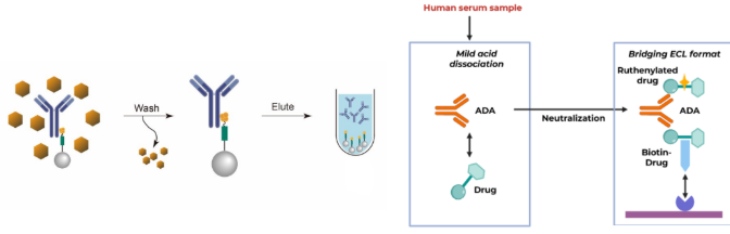
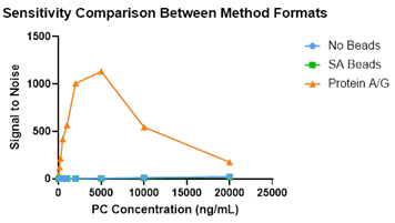
Because the transferred method lacked the required sensitivity for use in a clinical study, we evaluated alternative formats. A format of solution-phase acid dissociation followed by affinity capture elution (ACE) using biotinylated drug and streptavidin beads (SAB), like the original bridging method, showed very little specific signal. The format selected ultimately included a Protein A/G cleanup step to remove unrelated components from the sample.
This option was available because the drug is not an antibody and has no fragment antigen-binding (Fab) or fragment crystallizable (Fc) regions. Protein A/G therefore captures antibodies from the sample without co-eluting drug, both improving drug tolerance of the assay and removing excess FD1 from the assay system.
The final assay uses Protein A/G bead capture of antibodies from the sample, acid treatment to liberate the captured antibodies, and a standard master mix bridging assay. Detection used electrochemiluminescence on the Meso Scale Discovery (MSD) Sector Imager 600, leveraging the principle of signal being directly proportional to the magnitude of ADA
Comparing assay formats
| Format | Principle | Observation |
| Master Mix | Original transfer method, solution-phase master mix, no purification | Very little separation, sensitivity above 500ng/mL, very high background |
| Streptavidin bead ACE | Solution-phase acid capture and elution with biotinylated drug on magnetic beads | Very little separation, no improvement in sensitivity compared to original format |
| Protein A/G bead | Non-specific antibody capture, acid treatment, then bridging assay | Removed enough unrelated serum components to establish sensitivity |
Positive control selection
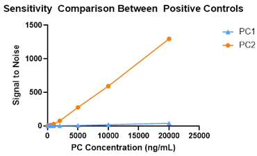
Three positive controls were screened: a rabbit polyclonal against the intact molecule, and two monoclonals specific to the FD1 moiety (positive control 1, PC1) or the FD2 moiety (positive control 2, PC2).
- PC1 (anti-FD1): not selective enough to act as a surrogate positive control. Homology of FD1 to a highly abundant protein in human blood impacted sensitivity due to cross-reactivity of PC1 with a native protein.
- PC2 (anti-FD2): allowed a sensitive curve, as no circulating target is expected in human blood
Validation outcome
- Screening, confirmatory and titer cut points were established.
- Sensitivity, drug tolerance and domain specificity met current FDA guidance and client needs
- The validated assay supported the Phase 1 trial (400 samples), and the Phase 2 trial is currently enrolling.
What made the difference
Protein A/G bead capture, an atypical approach for ADA assays, was successful for this non-antibody therapeutic. Our experience and expertise with ADA assay formats, assay condition optimization, and addressing sensitivity and drug tolerance challenges enabled the development of a validated method.
To know more about how we can support your immunogenicity programs, feel free to contact us here: