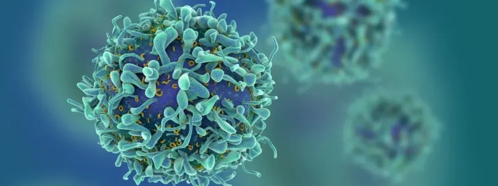
Many basic and clinical immunology studies that focus on T cells include proliferation assays in order to determine if T cells are capable of proliferating under different in vitro or in vivo conditions. Flow cytometry is the ideal approach for measuring T cell proliferation and a suite of staining products now exist that allow you to incorporate a proliferation dye into your existing flow cytometry staining panel. As with all flow cytometry staining protocols, be sure to do a pilot experiment to titrate your staining reagents and determine how many cells you need to run during your flow cytometry acquisition to have enough events for an accurate proliferation measurement.
Consider using one of these staining strategies to measure T cell proliferation in your next T cell assay.
Intracellular Fluorescent Staining:
Carboxyfluorescein diacetate succinimidyl ester (CFSE) or similar fluorescent stains are dyes that can be incorporated intracellularly into living cells. During proliferation, the fluorescent stain is diluted during subsequent cell divisions, and multiple rounds of cell division can be measured as discrete peaks during flow cytometry analysis. These proliferation dyes typically do not affect cell viability or morphology, but be sure to confirm this when you are testing such dyes in a pilot experiment.
DNA Synthesis:
Cell proliferation can be quantified by measuring DNA synthesis during the S phase of the cell cycle using a synthetic analog of thymidine called 5-bromo-2’-deoxyuridine (BrdU). BrdU is added in cell culture typically to determine in vitro proliferation, and BrdU incorporation is measured using intracellular cytokine staining with secondary antibodies against BrdU.
One of these T cell proliferation approaches may be more suitable to the type of experiment you are doing or the type of flow cytometer available to you. Each approach is relatively easy to add to your next experiment and can give you valuable insight into T cell function.
https://www.flowmetric.com/improve-preclinical-prospects-using-flow-cytometry-preclinical-development?hsCtaTracking=ff910900-4678-407b-b849-34bb090b52bf%7C2c6acede-4600-477c-8fb9-b1add09c33a6

