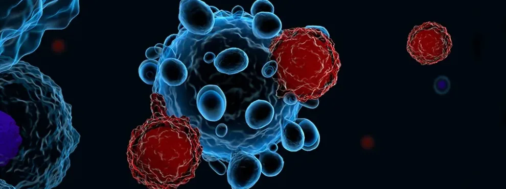
T cells are well known for their roles in combating cancer and infection, but chronic exposure to antigens and inflammation can cause T cells to enter a state of “exhaustion[1].” Exhausted T cells lose critical effector functions including cytokine production, the ability to proliferate and memory T cell differentiation is also compromised. Exhausted T cells also express inhibitory receptors and become unresponsive to IL-7 and/or IL-15-driven self-renewal. This progression toward T cell exhaustion results in diminished control of chronic infection or cancer. Exhaustion can occur in both CD4+ and CD8+ T cell populations and the phenotypes of these subsets is somewhat heterogeneous. Nonetheless, T cell exhaustion is reversible and various immuno-oncology interventions have been examined or are currently being evaluated in order to improve outcomes in cancer and chronic infection[2].
Using Flow Cytometry to Monitor Exhaustion
Customized flow cytometry panels can be created to monitor CD4+ or CD8+ T cell exhaustion, and the level of complexity of these panels is only limited by the type of cytometer available for use. Exhaustion of CD4+ T cells is typically associated with co-expression of multiple inhibitory receptors, including PD-1, CTLA-4 and LAG-3, as well as decreased production of cytokines such as TNF-alpha and IFN-gamma, and decreased proliferative and cytotoxic functionality in vitro. Alterations in transcription factor expression, including EOMES and GATA-3, can also be measured. CD8+ T cells also show increased co-expression of inhibitory receptors, impaired production of cytokines and diminished proliferative and cytolytic function. In addition, CD8+ T cells are less responsive to IL-7 and IL-15, which leads to decreased memory T cell proliferation. Flow cytometry can be used to measure all of these parameters in these T cell subsets.

Additional proteome and exome sequences can accompany flow cytometry analysis to identify other markers of exhaustion, but for most pre-clinical and clinical applications, flow cytometry analysis is a powerful, flexible and relevant tool for monitoring this immune state.
[1] Wherry EJ, Kurachi M. Molecular and cellular insights into T cell exhaustion. Nat. Rev. Immunol.. 2015;15(8):486-499.
[2] Nguyen LT, Ohashi PS. Clinical blockade of PD1 and LAG3 — potential mechanisms of action. Nat. Rev. Immunol. 2014;15:45–56.

