 Blogs
Blogs
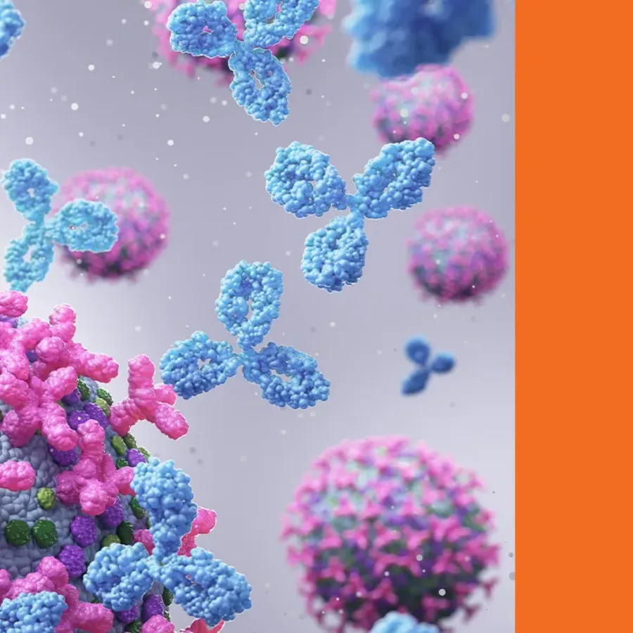
In the ever-evolving landscape of drug development, biomarkers have emerged as critical tools for the mechanism of action, early proof of mechanism, safety, predictive, efficacy, and monitoring treatment response. As the field advances, two major classes of biomarkers have come to the forefront: soluble biomarkers and cellular biomarkers. While…
 Blogs
Blogs
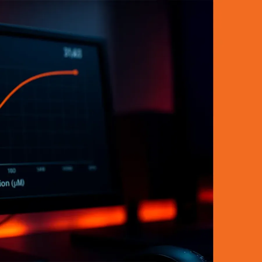
In the process of drug discovery, an IC₅₀ assay serves as a pivotal component in the development of cutting-edge therapeutics. An effective assay can quantify the potency of compounds, enable side-by-side comparisons with competing compounds, and track how a compound performs over time. Quantifying compound inhibition also assists with pre-clinical…
 Blogs
Blogs
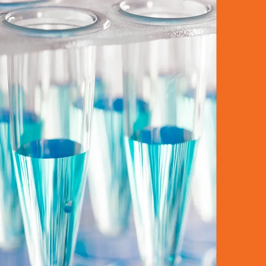
PCR-based assays are increasingly utilized for bioanalysis to support the development of a wide variety of therapeutics. While the largest driver for this growth has been the expanding pipeline of cell, gene, and RNA therapies, PCR-based assays are also seeing increased use in biomarker detection across all therapeutic modalities. These…
 Blogs
Blogs
Remember that childhood excitement the night before Christmas? Too excited to sleep, knowing something you wished for was finally within reach? That’s exactly the spirit fueling our teams in Lyon as we approach the end of June. We’re eagerly awaiting the arrival of a powerful new platform at our…
 Blogs
Blogs
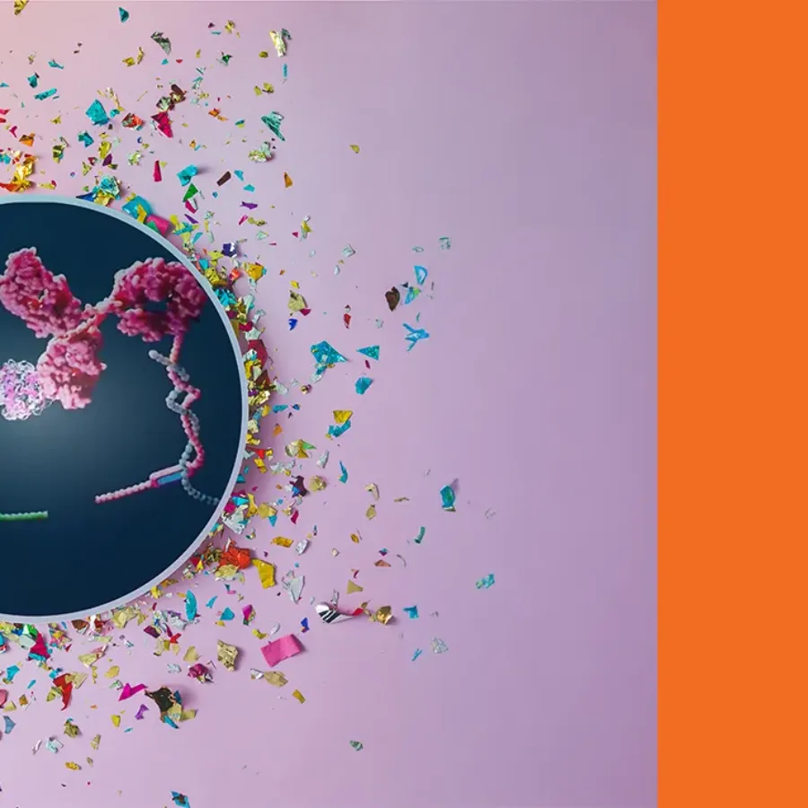
Hybrid LC-MS/MS is a technique that combines an affinity capture step with LC-MS/MS detection. It typically requires only one antibody, in contrast to conventional ligand-binding assays (LBAs), which usually need two. This approach leverages the combined selectivity of affinity extraction and the analytical power of tandem mass…
 Blogs
Blogs
Biomarkers (BMKs) have become fundamental tools in drug development, accelerating and optimizing targeted therapeutic innovation. As a bioanalytical CRO with over 15 years of experience in biomarker analysis, we’ve seen firsthand how the strategic integration of biomarkers can accelerate drug programs, help meet evolving regulatory standards,…
 Blogs
Blogs
From initial receipt to final disposition, sample integrity, traceability, and compliance are the foundation of our bioanalytical operations. We’re taking you behind the scenes to follow the journey of a sample through our facility. Here’s a detailed look at each stage in the lifecycle of a study sample at KCAS…
 Blogs
Blogs
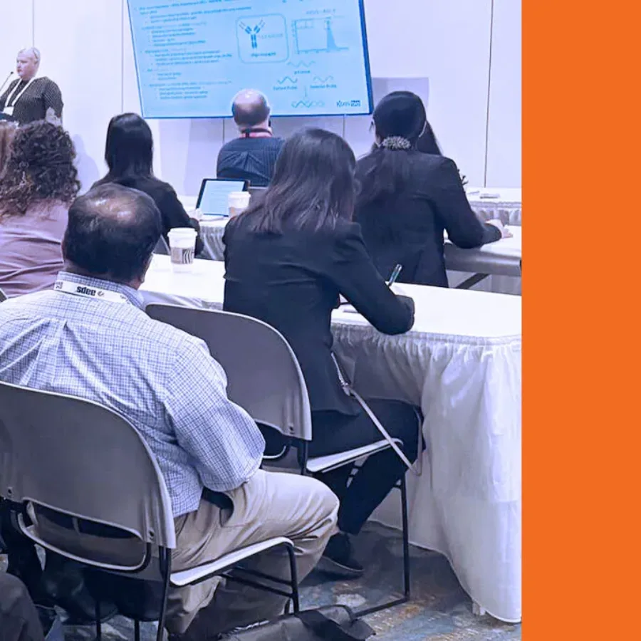
KCAS Bio participated in the AAPS National Biotechnology Conference (NBC), held from May 4 to May 7, 2025, in Boston. It was a productive event that allowed us to connect with sponsors, exchange ideas with peers, and present our latest work in bioanalytical science. Our experts…
 Blogs
Blogs
In drug development, the journey from target identification to clinical candidate selection involves a series of critical steps, including compound screening, lead optimization, and pre-clinical testing. Each stage helps narrow the pool of potential therapeutics, with the goal of identifying the most promising candidates for clinical evaluation. Given the time,…
 Blogs
Blogs
At KCAS Bio, we offer expert flow cytometry services designed to support the evolving bioanalytical needs of immunology and biomarker projects for drug development. Whether you’re working in preclinical or clinical settings, our validated off-the-shelf panels save time, reduce risk, and deliver high-quality, reproducible data you can trust. Extensive Portfolio…
 Blogs
Blogs
KCAS Bio offers a wide range of biomarker services, from cell-based to soluble biomarker analysis, including ligand binding assays (LBA), across a variety of matrix types. Soluble biomarker analysis can be achieved on multiple platforms depending on factors such as sample type, required sensitivity, and whether multiplexing is…
 Blogs
Blogs
Quantitative flow cytometry (QFCM) is a specialized technique that enables precise measurement of the absolute number of specific molecules (e.g., receptors, antigens, or intracellular targets) on individual cells or particles. Understanding flow cytometry is essential, as standard methods typically provide qualitative data, where the relative fluorescence intensity is used to…