How does flow cytometry work?
Flow cytometry is a powerful, high‑throughput technique that enables single‑cell analysis through fluorescent antibody staining, laser interrogation, and software‑driven gating workflows. At KCAS Bio, we guide your sample from preparation to data acquisition, analysis, and reporting, all under GLP/GCP‑compliant conditions.
Talk to an Expert

Flow Cytometry Process Overview
-
Step 1: Sample Preparation (Cellular)Cells from blood, tissue, or culture are suspended in fluid, filtered if needed, and stained with fluorescent antibodies targeting specific markers. Samples are then washed, possibly fixed, and stored to preserve viability and marker integrity.
-
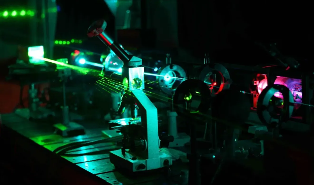
Step 2: Data AquisitionPrepared samples enter the flow cytometer, where cells pass single-file through a laser beam. Fluorescent signals and light scatter are detected in real time, generating raw data.
-
Step 3: Data AnalysisSoftware converts detector signals into metrics such as size, granularity, fluorescence intensity, and population frequency. Gating strategies define and evaluate specific cell subsets.
-
Step 4: ReportingResults are visualized with plots or tables, then compiled into reports for regulatory, scientific, or internal use.

Tell us how we can help with your project
We've earned our reputation for delivering reliable, error-free data. We understand the importance of speed, flexibility, and consistency and only make promises we can keep.
Talk to an expert