The Rise of Immune Cell Therapies and the Importance of Cell Profiling by Flow Cytometry
Immunotherapy is revolutionizing patient care, both in traditional oncology applications as well as in autoimmune indications. The complex mechanism of action based on the enhancement or targeted suppression of immune responses is fraught with challenges; as such it is critical to understand both the efficacy and safety of the pharmacodynamic profile of new interventional strategies. Development of these novel therapies therefore requires powerful immunophenotyping techniques and in-depth functional analysis to fully characterize the mechanism of action of the drug on biologically significant target cell populations.
In recent years flow cytometry has become a critical tool for drug developers who need to map complex mechanisms of immune perturbations. While flow-cytometry-based cellular phenotyping identifies immune subsets and their relative frequencies within a sample, functional assays allow for the deep characterization that point most directly to suppressive capabilities or activation status of a target cell population.
Neutrophils: An Important Yet Challenging Clinical Target
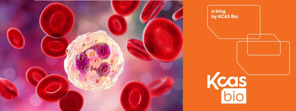
Neutrophils form the first line of defense against invading pathogens. These cells are rapidly recruited to the sites of infection. They destroy microorganisms by releasing reactive oxygen species (ROS) during the process of oxidative burst and engulfing pathogens by phagocytosis. The functional capacity of neutrophils to properly generate ROS and phagocytose pathogens is critical for the prevention of diseases such as Neutropenia and garners significant attention in cancer progression.
Neutrophil functional tests have proven to be complicated by ex vivo manipulations and mechanical processing. These cells are remarkably short-lived with a circulating half-life of 6-8 hours. Neutrophil phenotypes can be compromised simply by the required timeframe for sample processing. This can be observed by a varying level of activation and artificial changes, hence posing a challenge for accurate neutrophil characterization. Neutrophils require fresh processing and as such patient samples cannot be cryopreserved for delayed analysis. Oftentimes clinical samples are collected distant from the site of cellular analysis which creates delays in specimen handling. Neutrophil recovery and responses to stimuli can be easily affected by laboratory procedures and factors such as anticoagulant choice, incubation times, incubation temperatures, and a myriad of other laboratory handling issues.
Robust and Rapid Neutrophil Functional Phenotyping
Recently KCAS Bio has partnered with cutting-edge drug developers to implement functional neutrophil analysis assays as a critical part of clinical studies. This included neutrophil phagocytosis and oxidative burst potential analysis at different stages of cell maturation in whole blood.
The innovative aspect of the assays lies in incorporating quantification of neutrophil function with cellular phenotypic profiling in a single measurement in as little as 100 microliters of blood collected in sodium-heparin tubes. The assay design eliminates the need for long procedures such as neutrophil isolation which can lead to cell death in this extremely sensitive cell population.
The phagocytic activity of the cells is measured by cell-mediated ingestion of fluorescently labeled bacteria. As bacteria go through acidification during phagosome maturation the fluorogenic dye which is conjugated to bacteria dramatically increases in fluorescence as the environment pH becomes more acidic.
The oxidative burst (Oxiburst) assay uses an orthogonal functional strategy by introducing dihydrorhodamine 123, a non-fluorescent dye which is converted to fluorescent compound by reactive species produced by activated phagocytes when eliminating invading pathogens. The Oxiburst assay can be applied to any cell type which produces a NADPH oxidase-dependent respiratory burst response.
In the study method which will be featured at the upcoming 39th Annual Meeting Society for Immunotherapy of Cancer (SITC 2024), KCAS Bio qualified both assays and was able to demonstrate a stability of up to 24 hours. This stability is critical to supporting the transportation of samples from multiple clinical sites when executing a clinical study. The assays can be easily adapted to test other myeloid lineage cell populations such as macrophages. This recent development offers an innovative opportunity for rapid screening of therapeutic modalities targeting phagocytic cell deep functional phenotyping.
Unlock the Potential
KCAS Bio’s proven experience in developing neutrophil functional analysis assays can help unlock the full potential of your drug development. Our techniques ensure accurate, rapid phenotyping and functional assessment, even with the most sensitive cell populations. Reach out to us today to discuss how we can support your clinical studies and gain unparalleled insights into immune cell behaviors.

